Download
1 / 28
320 likes | 884 Views
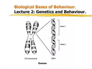
Biological Bases of Behaviour. Lecture 9: Cerebral Cortex. Kalat (2001) p 114. Learning Outcomes. At the end of this lecture you should be able to: 1 . Explain how the cortex is organised at a cellular level. 2 . Describe the gross anatomy of the cerebral cortex.

E N D
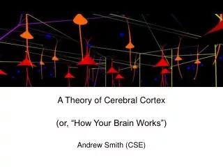
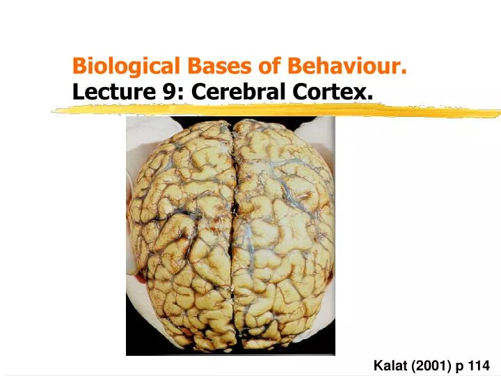
Biological Bases of Behaviour. Lecture 9:Cerebral Cortex. Kalat (2001) p 114
Learning Outcomes. • At the end of this lecture you should be able to: • 1. Explain how the cortex is organised at a cellular level. • 2. Describe the gross anatomy of the cerebral cortex. • 3. Describe the functional organisation of the 4 lobes. • 4. Outline the key behavioural features of the 4 lobes.
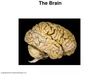
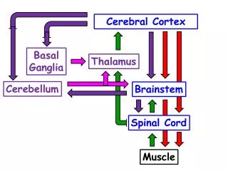
The Forebrain. • The forebrain consists of the two cerebral hemispheres. • Each hemisphere receives sensory information from the opposite (contralateral) side of the body, and controls the muscles on the contralateral side of the body. • The outer cellular layer of the hemispheres is called 'cortex' and consists of ‘gray matter’, axons descend from the cortex to form 'white matter'. • Hubel & Wiesel (1979): the cortex contains around 50-100 billion neurons, unfolded it would occupy an area of 2000cm² • Neurons in one hemisphere communicate with corresponding areas of the other hemisphere via two fibre pathways: the corpus callosum, and the anterior commissure.
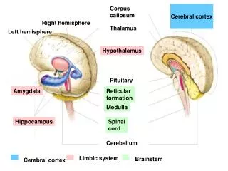
Key Features of the Forebrain. Longitudinal fissure Lateral ventricle Corpus callosum White matter Grey matter Central sulcus Anterior commissure Toates (2001) p 109
Cellular Organisation of the Cortex. • Cell structure and organisation varies between different regions of cortex. • These morphological differences relate to functional differences • The cortex contains up to six cell layers (laminae) separated by layers of fibres. • The laminae vary in density in different parts of the cortex, and not all layers are present throughout the cortex. Rosenzweig et al (1996) p63
Examples of Laminae Differences. • Layer IV contains small cells that receive sensory information and this layer is prominent in cortical regions which process sensory information. • Layer IV is absent in brain regions that control movement. • It is thicker in the visual cortex of people with photographic memories, and in the auditory cortex of musicians (Scheibel, 1984). • Layer V contains large pyramidal cells which are responsible for motor control. • Such cells predominate in areas of motor cortex.
Mapping the Cortex • Maps have been developed of cortical subregions based upon differences in cell density, cell shape, size, and connectivity. • Divisions based upon structural criteria define functional zones such as specialised areas for touch, perception and even distinct cognitive processes. Brodmann’s cytoarchitectonic map Kolb & Whishaw (1990) p 21
Columnar Organisation. • Cells that perform similar functions are organised into collumns each around 3mm deep, arranged perpendicular to the laminae. • E.g. if a single cell within a column responds to touch on the palm of the left hand, then other cells within the same column will also respond to that stimulus. • Mountcastle (1979) referred to these as macrocolumns and estimated that around a million of them existed in human cerebral cortex. • These can be further subdivided into minicolumns and there are an estimated half a billion of them.
Cortical Columns Surface of the cortex 3mm White matter Kalat (2001) p103
The 4 Lobes. • Regions of cortex are grouped together into 4 lobes named after the skull bones under which they lie: Parietal lobe Frontal lobe Occipital lobe Temporal lobe Kalat (2001) p104
1. Frontal Lobes. • These extend from the central sulcus to cover the anterior portion of the brain. • They contain: • i) Primary motor cortex (area 4). • ii) Premotor cortex (area 6). • iii) Broca's area (area 44). • iv) Prefrontal cortex. • Each receives input from the thalamic nuclei, limbic system, hypothalamus, and the other lobes, making it a 'control centre'. • Damage to the frontal part of the brain is thus likely to affect behaviour in many ways.
Divisions of the Frontal Lobes Primary motor cortex Premotor cortex Central sulcus Frontal cortex Prefrontal Cortex Broca’s area Goldman-Rakic (1984) p 10
Motor Cortex • Damage to the motor areas (4 and 6) produce a range of impairments to the motor system including: • i) Loss of fine motor control. • ii) Loss of strength. • iii) Interruption of open-loop motor programmes (sequences of fast muscle actions e.g typing, piano playing, speech). • iv) Loss of fine facial movements so patients with frontal lobe damage show relatively little spontaneous facial expression.
Broca's Area. • In 1861 Broca reported the case of a man who had lost the power of speech (though he could could still make speech noises and understand speech). • At autopsy the damage was found to be localised to a specific region on the left hemisphere of frontal cortex. • This impairment is now referred to as Broca's aphasia and is characterised by slow, deliberate speech with a very simple grammatical structure.
Prefrontal Cortex. • This forms around a third of the entire cortical mantle and constitutes a larger proportion of the brain in humans than in other species. Squirrel monkey Cat Rhesus monkey Dog Chimp Human Kalat (2001) p106
Role of Prefrontal Cortex. • A key role of prefrontal cortex concerns working memory - the ability to retain pieces of information for short periods of time (Goldman-Rakic, 1984). • Brain imaging studies, case studies of brain-damaged humans, single-cell recordings confirm that this region is extremely active during delayed response tasks. • Prefrontal cortex is also involved in higher-order cognitive behaviours: • Planning. • Organisation. • Monitoring events, their outcomes, and the emotional value of such actions.
Impairments Following Frontal Lobe Damage. • Temporal sequencing: i.e the ability to say which of 2 pictures has been presented most recently. • Shifting of attention: An increased tendency to persevere with an action when it is incorrect (perseveration). • Conditional associations: the ability to associate a correct response with a particular stimulus. • Working memory: the ability to maintain a response in memory and then act upon it appropriately. • Response inhibition: Cannot inhibit responses, take more risks, and demonstrate antisocial, aggressive, and sexually inappropriate behaviours. Don’t behave appropriately in certain situations (e.g. laughing during a funeral).
2. Temporal Lobes. • The temporal lobes comprise the tissue below the lateral fissure running posterior to parietal and occipital cortices. • They are described by gyri that form them - the superior temporal gyrus, the middle temporal gyrus, and the inferior temporal gyrus. • The temporal lobes are richly connected to the other lobes, the sensory systems, the limbic system, and the basal ganglia. • They seem to have 3 key functions: Kolb & Whishaw (1990) p 441
Functions of the Temporal Lobes. • i) Auditory and visual perception: Areas 22, 41 and 42 are concerned with focussing attention on relevant auditory information and with the perception of music and speech. • Damage to area 22 produces Wernicke's aphasia - the inability to understand words, or to arrange sounds into coherent speech despite having intact hearing. • Damage to the equivalent areas in the right temporal lobe produces deficits in processing music. • Right temporal lobe damage seems to produce a specific impairment in the recognition and recall of familiar faces (called prosopagnosia). • Patients also have difficulty perceiving subtle social signals. • Tumours or epilepsy confined to the temporal lobes often give rise to auditory or visual hallucinations.
ii) Memory. • The case of HM ( Scoville & Milner, 1957) showed that the hippocampus is critical for laying down new memories. • Following radical temporal lobe surgery for intractable epilepsy, HM is unable to form new memories (anterograde amnesia) Destroyed hippocampus in patient HM Kalat (2001) p371
iii)Emotion and Mood. • The amygdala is concerned with emotional control. When exposed to photographs displaying emotional expressions (especially fear) the amygdala is strongly activated. • Patients with amygdala damage do not show appropriate responses to fear-inducing stimuli nor can they recognise emotional expressions on other people’s faces. Hippocampus Amygdala Kolb & Whishaw (1990) p 442
3. Parietal Lobes. • These lie between the occipital lobe and the central sulcus. Behind the central sulcus lies the postcentral gyrus housing primary somatosensory cortex (the sensory representation of the body). Kalat (2001) p105
The Sensory Homunculus. • The right hemisphere contains information about the left side of the body and vice versa. • Some body parts (hands, fingers, lips, genitals) receive more regions of cortex devoted to their sensory representation. • Experience can modify these cortical representations. • E.g. string musicians show an enlarged representation of the fingers of the left hand in the right hemisphere somatosensory strip (Kalat, 2001).
Damage to the Parietal Lobes. Patient’s response target • Damage here produces deficits in: • Tactile function. • Disorders of body image. • Right-left confusion. • Disorders of spatial ability (Kolb & Whishaw, 1990). • A common feature is sensory neglect, the tendency to ignore one side of the body or features of the outside world. Kolb & Whishaw (1990) p 425
4. Occipital Lobes. • Occipital cortex comprises primary visual cortex (striate cortex or V1) and visual association areas (V2-V5). • Damage to occipital cortex produces many deficits of visual perception including blind spots in the visual fields (scotoma), dyslexia, colour blindness and problems in perceiving faces or objects (agnosia). Rosenzweig et al (1996) p349
The ‘Binding Problem’. • We perceive an integrated world despite the fact that neural processing is conducted by distinct (but interconnected) modules. • How are separate functions integrated? • As yet this remains a mystery but Robertson et al., (1997) proposed that regions of inferiorparietal cortex may serve to combine different aspects of information to form a coherent whole. • This theory is based upon individuals with brain damage to parietal cortex who can no longer bind together different aspects of perception.
References and Bibliography. • Goldman-Rakic, P.S. (1984). The frontal lobes: uncharted provinces of the brain. Trends in Neurosciences, November: 7-11. • Hubel, D.H., & Wiesel, T.N. (1979). Brain mechanisms of vision. Scientific American, 241: 150-168. • Kalat, J.W. (2001). Biological psychology. • Kolb, B., & Whishaw, I.Q. (1990). Fundamentals of Human Neuropsychology. • Mountcastle, V.B. (1979). An organizing principle for cerebral function: the unit module and the distributed system. In F.O.Schmitt and F.G.Worden (Eds). The neurosciences, MIT Press, pp 21-42. • Robertson, L., Triesman, A., Friedman-Hill, S., & Grabowecky, M. (1997). The interaction of spatial and object pathways: evidence from Balint's syndrome. Journal of Cognitive Neuropsychology, 9: 295-317. • Rosenzweig, M.R., Leiman, A.L., & Breedlove, S.M. (1996). Biological Psychology.
References and Bibliography. • Scheibel, A.B. (1984). A dendritic correlate of human speech. In N.Geschwind and A.M.Galaburda (Eds.), Cerebral dominance, Harvard University Press, pp 43-52. • Scoville, W.B., & Milner, B. (1957). Loss of recent memory after bilateral hippocampal lesion. Journal of Neurology, Neurosurgery and Psychiatry, 20: 11-21. • Toates, F. (2001). Biological psychology an integrative approach. Prentice Hall, chapter 5.