aorta
200 likes | 689 Views
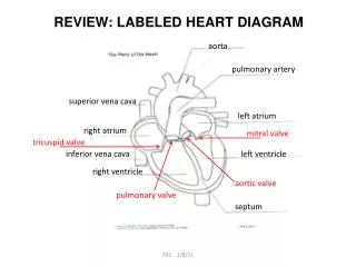
REVIEW: LABELED HEART DIAGRAM. aorta. pulmonary artery. superior vena cava. left atrium. right atrium. mitral valve. tricuspid valve. inferior vena cava. left ventricle. right ventricle. aortic valve. pulmonary valve. septum. The Heart Beat.

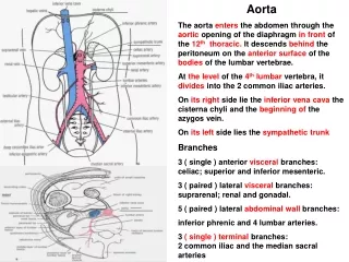
aorta
E N D
Presentation Transcript
REVIEW: LABELED HEART DIAGRAM aorta pulmonary artery superior vena cava left atrium right atrium mitral valve tricuspid valve inferior vena cava left ventricle right ventricle aortic valve pulmonary valve septum 392 2/8/11
The Heart Beat Systole - Heart is pumping blood, contracting muscle • atria to the ventricles • ventricles to body or lungs Diastole – Heart is relaxing • atria fill with blood How can the heart be monitored for normal activity? 392 2/8/11
The EKG (or ECG)Electrocardiogram • Measures the electricity passing through the heart at a given time • Can be used to diagnose heart conditions • Each section of the EKG shows a specific event happening to the heart. 392 2/8/11
The EKG Tracing P wave = atrial contraction QRS peak = ventricle contraction T wave = resetting of the heart systole diastole 392 2/8/11
Circulation Through the Heart 392 2/8/11
TWO CIRCULATORY PATHWAYS Capillaries of head and arms Superior vena cava Pulmonary artery Aorta PULMONARY CIRCULATION Pulmonary vein Capillaries of left lung Capillaries of right lung SYSTEMIC CIRCULATION Inferior vena cava Capillaries of abdominal organs and legs 392 2/8/11
Blood Vessels 392 2/8/11
Endothelium Arteriole Venule Connective tissue Connective tissue Smooth muscle Smooth muscle Endothelium Endothelium Valve Structure of Blood Vessels Artery Vein Capillary 392 2/8/11
Cross Section Through Blood Vessels 392 2/8/11
Arteries carry oxygen-rich blood away from the heart to the body's tissues. Arteries carry large quantities of blood that is under high pressure from the beating of the heart, they are wide and thick. Capillaries are very narrow -- only one cell wide. They have very thin walls made of overlapping flat cells called endothelium; the walls are thin so that oxygen and carbon dioxide can pass through them easily. Veins take oxygen-poor blood back to the heart. From the capillaries, the de-oxygenated, waste-laden blood passes into the veins for its return trip to the heart. Veins contain one-way valves to keep the blood flowing toward the heart. 392 2/8/11
Blood Pressure (BP) • What is it? • The force of the blood against the walls of the arteries • What does a blood pressure reading mean? • Blood pressure is recorded as two numbers, written as a ratio. For example, • 120/80 mm Hg • Top number is SYSTOLIC BP, pressure in arteries when the heart contracts. • Bottom number is DIASTOLIC BP, pressure in arteries when heart rests 392 2/8/11
Blood Pressure versus Heart Rate • Heart Rate is the number of times your heart beats per minute; your PULSE. • There is no good connection between heart rate and blood pressure. • For cardiovascular health, KNOW YOUR BP! 392 2/8/11