Download
1 / 13
180 likes | 894 Views
Benign Mucous Membrane Pemphigoid. Kelly Shepard Oral Medicine and Diagnosis Case Study UMKC School of Dentistry Fall 2010. UMKC Dental School Patient Record # 520-8-08. Patient Background. 86 Year-Old, Caucasian Female Brought into the office for Routine Dental Care

E N D
Benign Mucous Membrane Pemphigoid Kelly Shepard Oral Medicine and Diagnosis Case Study UMKC School of Dentistry Fall 2010
Patient Background • 86 Year-Old, Caucasian Female • Brought into the office for Routine Dental Care • Medical History Reveals: • Arthritis, Back pain, Controlled Hypothyroidism • Medications: • Meloxicam, Simvastin, Synthroid • Regularly sees physician and optometrist • No complaints of tooth or periodontal related discomfort • Psychologically: patient is hesitant to disclose or acknowledge medical history
Intraoral PHotographs UR arrow: Lesion after confirming a Positive Nikolsky Sign
Lesion Description • Predominately found on the mucous membranes of the oral mucosa this patient has several intraoral bullae or vessicles that range in size from 3 mm to 2 cm. • The underlying ulcers are symmetric, well defined, and are surrounded by a red area of inflammation. • The lesion appears to involve the epithelium. • Patient presents with generalized desquamative gingivitis. • The patient has several other lesions throughout the mouth and on the attached gingival and mucosal tissue.
Patient Dialogue Have you ever noticed these lesions before? Only after the bridge was cemented Has your eye doctor or physician ever diagnosed you or a family member with a vesibulobulous disease? No Do you have any other lesions such as this anywhere else on your body? No Do these ever cause you any discomfort? Patient claims these lesions were caused by the nearby bridge and has noticed discomfort with them since then. When was the last time you were seen by your optometrist? 6 months ago (patient was not aware if it was an optometrist or ophthalmologist)
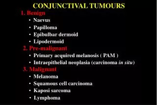
Differential Diagnosis Mucous Membrane Pemphigoid Common chronic disease involving patients over 50 years old, affecting the oral cavity, genitals, and conjunctiva PemphigusVulgaris Fatal bullous disease of the skin and mucosa, associated with painful vessicles and shallow ulcers Erosive Lichen Planus Commonly associated with desquamative gingivitis Erythemamultiforme Acute vesicular-bullous disease characterized by rupturing vesicles and bulla with bleeding ulcerations Definitive Diagnosis pending Biopsy: Mucous Membrane Pemphigoid
Additional Lesions Symblepharons: Ocular lesions associated with Mucous Membrane Phemphigoid. These are epithelial adhesions between the bulbar and palpebral conjunctivae. Blindness occurs in 1/3 of patients.
Recommended Treatment: • Definitive Diagnosis: light microscopy & direct immunofluorescense • Referral: Opthalmologist or other Specialist as needed • Topical Medicaments • Daily Topical corticosteroids until resolved • Resume as lesions continue to flare up • Flexible mouth guard as a corticosteroid carrier • Systemic Medicaments • Immunosuppresive agents • IV human immunoglobulin • Antibiotics • Good Oral Hygiene • Due to the immunological involvement of the disease no cure or active treatment is available, medications are given simply in order to control the pain.
Patient Follow Up • Patient denied biopsy • Patient was not interested in treating the lesion • Patient has not returned for routine dental care due to back pain/arthritis • Patient states she has an appointment with an optometrist in December
References • Moghadam, Behjat. Oral Medicine and Diagnosis, Course Notes 2010 • Langlais et Al, Robert. Color Atlas of Common Oral Diseases. 4th Edition. Philadelpia, PA: Lippincott Williams and Wilkins, 2009. • Neville, Oral and Maxillofacial Pathology. 3rd. St. Louis, Missouri: Saunders: Elsevier, 2009.
Additional Photos UL/LL photo: desquamativegingiva UR: tissue response to air LR: additional lesion