Download
1 / 1
10 likes | 66 Views
Optical Science. Laboratory. Abstract.

E N D
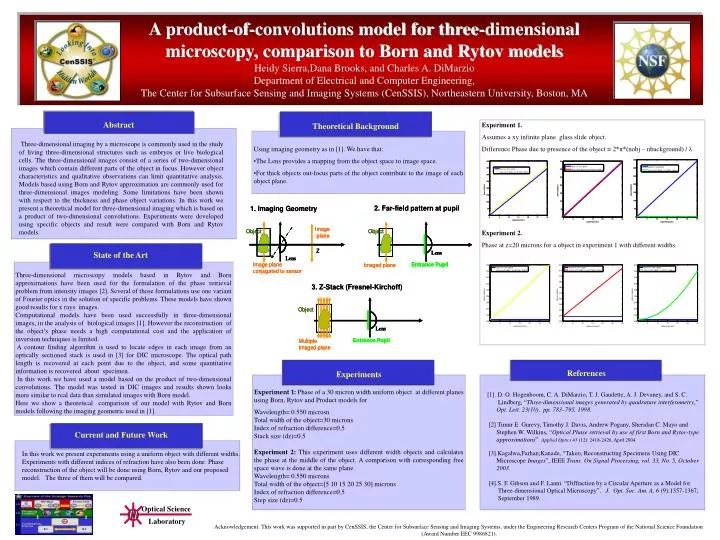
Optical Science Laboratory Abstract Three-dimensional imaging by a microscope is commonly used in the study of living three-dimensional structures such as embryos or live biological cells. The three-dimensional images consist of a series of two-dimensional images which contain different parts of the object in focus. However object characteristics and qualitative observations can limit quantitative analysis. Models based using Born and Rytov approximation are commonly used for three-dimensional images modeling. Some limitations have been shown with respect to the thickness and phase object variations. In this work we present a theoretical model for three-dimensional imaging which is based on a product of two-dimensional convolutions. Experiments were developed using specific objects and result were compared with Born and Rytov models. State of the Art Current and Future Work Experiments References A product-of-convolutions model for three-dimensional microscopy, comparison to Born and Rytov modelsHeidy Sierra,Dana Brooks, and Charles A. DiMarzioDepartment of Electrical and Computer Engineering,The Center for Subsurface Sensing and Imaging Systems (CenSSIS), Northeastern University, Boston, MA Theoretical Background Experiment 1. Assumes a xy infinite plane glass slide object. Difference Phase due to presence of the object = 2*π*(nobj – nbackground) / λ Experiment 2. Phase at z=20 microns for a object in experiment 1 with different widths. • Using imaging geometry as in [1]. We have that: • The Lens provides a mapping from the object space to image space. • For thick objects out-focus parts of the object contribute to the image of each object plane. Three-dimensional microscopy models based in Rytov and Born approximations have been used for the formulation of the phase retrieval problem from intensity images [2]. Several of those formulations use one variant of Fourier optics in the solution of specific problems. These models have shown good results for x rays images. Computational models have been used successfully in three-dimensional images, in the analysis of biological images [1]. However the reconstruction of the object’s phase needs a high computational cost and the application of inversion techniques is limited. A contour finding algorithm is used to locate edges in each image from an optically sectioned stack is used in [3] for DIC microscope. The optical path length is recovered at each point due to the object, and some quantitative information is recovered about specimen. In this work we have used a model based on the product of two-dimensional convolutions. The model was tested in DIC images and results shown looks more similar to real data than simulated images with Born model. Here we show a theoretical comparison of our model with Rytov and Born models following the imaging geometric used in [1]. Experiment 1: Phase of a 30 micron width uniform object at different planes using Born, Rytov and Product models for Wavelength= 0.550 microsn Total width of the object=30 microns Index of refraction difference=0.5 Stack size (dz)=0.5 Experiment 2: This experiment uses different width objects and calculates the phase at the middle of the object. A comparison with corresponding free space wave is done at the same plane. Wavelength= 0.550 microns Total width of the object=[5 10 15 20 25 30] microns Index of refraction difference=0.5 Step size (dz)=0.5 [1]. D. O. Hogenboom, C. A. DiMarzio, T. J. Gaudette, A. J. Devaney, and S. C. Lindberg, “Three-dimensional images generated by quadrature interferometry,” Opt. Lett. 23(10), pp. 783–795, 1998. [2] Timur E. Gurevy, Timothy J. Davis, Andrew Pogany, Sheridan C. Mayo and Stephen W. Wilkins, “Optical Phase retrieval by use of first Born and Rytov-type approximations”. Applied Optics 43 (12): 2418-2426, April 2004. [3].Kagalwa,Farhan;Kanade, “Takeo, Reconstructing Specimens Using DIC Microscope Images”, IEEE Trans. On Signal Processing, vol. 33, No. 5, October 2003. [4].S. F. Gibson and F. Lanni. “Diffraction by a Circular Aperture as a Model for Three-dimensional Optical Microscopy”. J. Opt. Soc. Am. A, 6 (9):1357-1367, September 1989. In this work we present experiments using a uniform object with different widths. Experiments with different indices of refraction have also been done. Phase reconstruction of the object will be done using Born, Rytov and our proposed model. The three of them will be compared. Acknowledgement: This work was supported in part by CenSSIS, the Center for Subsurface Sensing and Imaging Systems, under the Engineering Research Centers Program of the National Science Foundation (Award Number EEC 9986821).