Download
1 / 29
340 likes | 613 Views
Hoof Dissection. Equine Hoof Anatomy 101 Presented by Nancy Frishkorn AA, BA CHCP Liberated Horsemanship . Duckett’s Dot. Developed 28 years ago by British farrier David Duckett FWCF (resides in Pa. USA) Points on the horse’s foot ‘least likely’ to change

E N D
Hoof Dissection Equine Hoof Anatomy 101 Presented by Nancy Frishkorn AA, BA CHCP Liberated Horsemanship
Duckett’sDot • Developed 28 years ago by British farrier • David DuckettFWCF (resides in Pa. USA) • Points on the horse’s foot ‘least likely’ to change • Attempted to designate center of P3 • Total hoof length/2 = true apex • Approx 3/8” behind is the “DOT” 3/8” more “Bridge” • Claim was that Bridge is center of weight bearing area • Apex length/2 designated tip of P3 placement • Challenged by Mark Caldwell FWCF • Instructor at MyerscoughCollege in England • Recently received PhD- Distal Limb Morphometrics • University of Liverpool Department of Veterinary Clinical Science in Liverpool Nancy Frishkorn http://www.hooflady.org
Comparing“mappinglogic?” by Mark Caldwell Elemental discussion at the Northeast Association of Equine Practitioners Symposium Sept. 2012 in Pa. Nancy Frishkorn http://www.hooflady.org
Mapping…A Waste of Time! These are all living horses That I encountered and Healed with NHC! Nancy Frishkorn http://www.hooflady.org
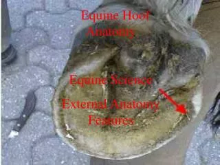
The Primary Hoof Structure The hoof has 5Coriums, or areas of the hoof that are named for the epidermal or horny structure for which they support. The dermis is similar to the quick on your fingers and the epidermis is like your skin and nails. Nancy Frishkorn http://www.hooflady.org
Notching Toe Wall • Toe wall rasped/nippered back to P3 • Laminaebreached well before indicated marking from Duckett’s “DOT” • Measurements did not prove accurate • Mapping of no use in deformed hooves • Squaring or backing toe wall not advised • Water line indicates inner 1/3 of hoof wall • Rolling only to the water line • will assure safe wall removal • Sole plane is best indicator for Toe Length Nancy Frishkorn http://www.hooflady.org
Sole Removal After slicing the hoof with a band saw, the Sole Corium can be easily peeled away from the hard sole plane, consequently revealing the Frog Corium as a separate entity from the Frog Stay (horny frog). Here is the junction between the sole and the inner coronary band that is not visible, as it lies under the sole plane. Nancy Frishkorn http://www.hooflady.org
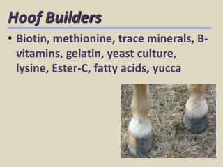
Sole Thickness • The Solar Corium attaches to the pedal bone from the toe • to the underside of the digital cushion. • Arranged into papillae • Sole Coria and Frog Coria merge together • Degree of concavity varies in every equine • Depends upon shape of coffin bone • Arched structure more important than concavity itself • Link to Ann Corso’s article “Form Follows Function” • Typical thickness is approx. 3/8 inch • Trim only to hard sole plane, never to solar corium • Natural concavity forms naturally, it is never “carved” Nancy Frishkorn http://www.hooflady.org
Dermal Laminae • Approx. 600 interlocking Dermal and Epidermal Laminae • Suspend and attach the coffin bone inside the hoof capsule • Each Dermal Laminaeslots in between epidermal leaves • This is sometimes called the “sensitive” laminae • Surface corrugations called Periosteum increase area of attachment • Periosteum contains arteries, veins and nerves • Small corrogations called Desmones microscopically visible • Hemodesmonesare contained within Desmones • Supply blood to lamellar leaves • Bar Laminae are visible on the underside sole corium • They are an extension of the coronary corium • Combined they create the Heel Buttress Nancy Frishkorn http://www.hooflady.org
Epidermal Laminae • Hemodesmones are supported by a thin lining membrane • These basal cells produce horny Epidermal Laminae • The membrane is termed Basement Membrane • Basal cells divide by mitosis • They “Keratinize” or harden to become Epidermal Laminae • Damage or expose of Dermal Membrane can lead to “Cornification” • Keratinized (Cornified) laminae are not sensitive • Horse shoe nails are driven through epidermal laminae • Invasion of dermal laminae can lead to abscess • Horse may be lame or have “hot spot” • Typically referred to as “hot nail” • Digital thermometer can detect damaged area • PULL THE SHOES to permit • healingof the capsule Nancy Frishkorn http://www.hooflady.org
The Coronary Band • The Coronary Band is a circular organ • fingerlike projective dermal papillae • Surrounds the hoof capsule joining horn to hairline • Lies under the digital cushion as frog corium • Looks like “Swiss cheese” and is filled with holes • Each hole or pit contains one Coronary Papilla • Each Papilla is responsible for producing one Horn Tubule • Each tubule has a hollow core called a Medullae • Each Tubular horn is separated by these inter-tubular horn • Coronary Corium (CC) produces the bulk of the hoof wall • Perioplic Corium lies above CC • Produces soft thin layer of tubular horn (periople) • Rarely extends more than 1” down hoof wall Nancy Frishkorn http://www.hooflady.org
Digital Cushion • The Digital Cushion lies underneath the frog and frog “stay” • It’s primary purpose is shock absorption • Protects the DDFTand Navicular bone • Does not “toughen” or become fibrocartilage • Increased movement aids in health and production • Feel is similar to “chicken fat” • Light and porous material • Extends into area of bulbs at back of foot • Flattens under weight to disperse shock • Dual purpose-as it flattens it pushes lateral • cartilages outward which expands the capsule Nancy Frishkorn http://www.hooflady.org
The Tendons • Tendons are bands of fibers that connect muscle to bone • Each tendon has “check” ligaments that keep it from overstretching • Two classifications • Extensors-Straighten the limb • Flexors-Bend the limb • Tendons are surrounded by synovial membrane (sheath) • There are no muscles in the lower limbs of equuscallibus • The Radius and Ulna bones in the upper leg have muscle • These connect to the Carpals • Called Carpi Radials • FaciaLatae/Bicep Femoris muscles in hinds Nancy Frishkorn http://www.hooflady.org
ExtensorTendon • Large tendon running down front of leg • Common Digital Extensor • Attached to (is part of) CDE muscle • Located from bottom 1/3 of radius to coffin joint • Lateral Digital Extensor • Attached to (is part of) LDE muscle • Inserts into the proximal portion of the first phalanx • Extends the fetlock, pastern and coffin joints • Extensor Tendon of the Knee (Carpus) • Lies just above the knee joint of carpus • Straightens out the knee • Commonly injured due to striking against wall or post Nancy Frishkorn http://www.hooflady.org
FlexorTendon • Two Digital Flexor Tendons –Superficial and Deep • Superficial Digital Flexor (pictured to left) • Forms rear outline of the leg • Most frequent injury site resulting in Tendonitis • Often occurs when muscles over fatigued • Deep Digital Flexor Tendon (DDFT) detail next slide • In the hind limb the flexors straighten the hock • Achilles Tendon is visible as ridge in hind hock/leg • Comprised of several tendons including digital flexor • Peroneus Teritusis also part of the hind apparatus • Automatically bends the hock when the stifle is flexed Nancy Frishkorn http://www.hooflady.org
Deep Digital Flexor Tendon (DDFT) • Lies beneath the Superficial Flexor Tendon • Found in both the fore and hind limbs • Extends under the coffin bone between Palmer Process • Enters (connects) to P3 just behind the frog apex • Acts as a pulley system for the navicular bone • Simultaneously moves with action of superficial tendon • Both tendons are surrounded by the Annular Ligament • Sheath that forms a canal though which they pass freely • A tear or cut to this tendon causes fetlock to drop • Toe will tilt up under weight bearing Nancy Frishkorn http://www.hooflady.org
TheTenotomy • Achillistenotomies in humans are used to “correct” clubfoot • Veterinary community viewed founder as tendon related • Belief was that tendon over contracted • Severing tendon would stop rotation of P3 • “surgeries performed at mid cannon level resulted in • 6/20 cases surviving longer than 6 months” • Hunt, RJ. et al Vet Surg 1991;20:15–20. • “We measured success as the ability to survive 2 • years or greater….a 60% success rate seems encouraging, • especially considering the lack of available alternatives” • Texas A&M Eastman, T., et al AAEP PROCEEDINGS Vol. 44 / 1998 • 19 of 37 horses survived for breeding use or pasture • ornamentation...yet this was seen as a success-- • NHC IS THE ONLY ALTERNATIVE! Nancy Frishkorn http://www.hooflady.org
The Bones Within Nancy Frishkorn http://www.hooflady.org
Palmer Process • The Palmer Process is a cartilage material (wings) • CallateralCartliageslie atop the palmer process • Inside are the Digital Arteries (DA) • DA enter P3 through two holes just in front of DDFT • Inside they form a semicircular “Terminal Arch” • 9 branches pass through Pedal bone rejoining to exit • between P3 and the sole as the “Circumflex Artery” • The DA’s run from the heels along the frog sulci, • to the frog apex • No arterial branches are under the frog • No blood vessels are present in the sole Nancy Frishkorn http://www.hooflady.org
NavicularPlacement • The Navicular is suspended with 3 Impar Ligaments • Located at the heel (central 1/3 of frog) • Support for P3 and short pastern bones • It guides the DDFT into entry point • Lies under end point of DDFT • Navicular Bursa most proximal point • Entire structure called navicular complex • Injury to P3 can lacerate ligament • Resulting in upward • migration of Navicular bone Approx. frog location (DDFT removed) Nancy Frishkorn http://www.hooflady.org
Palmer View Navicular/P3 Here you can see how the navicular bone fits snugly into the back of the lowest phalanges. It is enclosed by the wings of the coffin joint (palmer process) and serves as a pulley for the deep digital flexor tendon. The navicular bursa is a sac that has a membrane lining to secrete lubricating fluid (synovial), and is located behind the navicualr bone. Collateral and bone erosion can occur resulting in degenerate changes and navicular syndrome. Proper, NATURAL hoof care, frequent trimming, exercise and dietary management all lesson the probability of developing navicular syndrome. Nancy Frishkorn http://www.hooflady.org
NavicularBone Nancy Frishkorn http://www.hooflady.org
SesamoidBones • Three sesamoid ligaments run between bones • Connect to back of short pastern and P3 • Comprise the suspensory apparatus (1-3) of fetlock • Suspensory ligament (wide/thick)extend • from lower cannon bone to upper long pastern • They divides (bifurcate) over sesamoid bones • Paired Sesamoid bones & ligaments • Superficial & Deep flexor tendons • Primary Purpose • Prevent overextension of fetlock joint • Absorb shock/concussion • Support the fetlock Nancy Frishkorn http://www.hooflady.org
Coffinbone Entry points for Digital Arteries In front of DDFT just behind apex (apex marked by screw) Extreme close up of the normal porous (vein/artery filled) surface Nancy Frishkorn http://www.hooflady.org
Short Pastern/Coffin Bone Nancy Frishkorn http://www.hooflady.org
Long Pastern Bone P1 P2 P3 Nancy Frishkorn http://www.hooflady.org
Cannon& Splint Bones Nancy Frishkorn http://www.hooflady.org
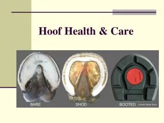
Hoof with Wall Removed So now you know some of the basic anatomy of the equine hoof and lower limb…when you look at the hoof , try to see the structures that lie just below the surface. Always respect the healing powers of nature and never violate these sensitive internal functions-least you hinder the healing process. When in doubt, DON’T MAP IT! Consult with your veterinarian and get x-rays… NO ONE has x-ray vision, and this is the ONLY way to know what’s TRULY going on inside the hoof! Nancy Frishkorn http://www.hooflady.org
References: All of the information was provided through my training programs with Liberated Horsemanship and the AANHCP. Personal notes and photos were used from my own collection, aside from the two hoof mapping images; these can be found in the links below. I sincerely hope you enjoyed my presentation and I look forward to instructing each and every one of you in either the field or at your barn! Daily rates for my instruction services can be found and paid for through Liberated Horsemanship at http://liberatedhorsemanship.com/Faculty.html Duckett’s Dot Mark Caldwell Nancy Frishkorn http://www.hooflady.org