Download
1 / 12
120 likes | 229 Views
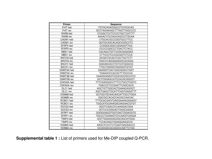
Supplemental table 1 : List of primers used for Me-DIP coupled Q-PCR.

E N D
Supplemental table 1 : List of primers used for Me-DIP coupled Q-PCR.
Supplemental table 2. 77 genes reported to be methylated in cervical neoplasia with a supporting PMID reference number; 1 identifies those genes with increased methylation following transfection of PHFK with episomal HPV16; 2 those with increased methylation following HPV18 transfection; and 1,2 those hypermethylated by both HPV types. Of 5837 genes with increased methylation in HPV16 or HPV18 transfected cells, 35 (0.6%) were also aberrantly methylated in cervical neoplasia as were 42 (0.3%) of those 14048 genes which were not hypermethylated following transfection with these high risk HPV types.
A B C A B C A B C A B C A B C A B C A B C A B C A B C Supplemental figure 1. Changes in methylation status detected using Me-DIP coupled Q-PCR in 3 additional PHFK donors (A, B and C) following transfection with episomal HPV18.
A B Supplemental figure 2. Simulation analysis designed to determine whether the observed frequency of concordant methylation changes in adjacent genes could result from a random distribution of HPV-induced methylation changes. Following the exclusion of 1596 genes with over-lapping transcriptional start sites; 5188 genes were found to have increased methylation following HPV16 transfection of which 3336 were located immediately adjacent to another hypermethylated gene; of 3290 genes with decreased methylation in HPV16 transfected cells, 1622 were immediately adjacent to another hypomethylated gene. Of 2599 genes found to have increased methylation following HPV18 transfection, 1305 were located immediately adjacent to another hypermethylated gene; of 3822 genes with decreased methylation in HPV18 transfected cells, 2025 were immediately adjacent to another hypomethylated gene. Histograms show the distribution of the number of adjacent pairs in 100000 simulated gene sets each containing 5188 genes (Panel A), 3290 genes (Panel B), 2599 genes (Panel C) and 3822 genes (Panel D) randomly selected from the total of 18389 genes. C D
A B C D Supplemental table 3. Panel A lists HPV16 and HPV18 hypermethylation and hypomethylation hotspots. Panel B lists those loci of HPV integration reported in the literaturewhich could be mapped onto the promoter array; each locus is supported by a reference (301, 312, 323, 334, 345). Panel C and D lists those regions of chromosomal loss and gain reported in Wilting et al., 2007 (37) which could be mapped onto the promoter array.
Supplemental table 4. Change in methylation status following transfection of PHFK with episomal HPV18 is related to promoter CpG content and to the pattern of histone marking in human embryonic stem (HES) cells. Risk of increased methylation increases with increasing CpG content and is greatest for genes bivalently marked by H3K4me3 and H3K27me3 in HES cells; risk of decreased methylation is lowest for bivalently marked genes. *Analysis restricted to genes listed on Weber et al., array (2007) (42). **Analysis restricted to genes listed on Zhao et al., array (2008)(47).
Supplemental table 5. Age-related polycomb target genes which are significantly more likely to be methylated in cervical smears taken from women with HPV associated CIN than in normal cervical cells and to have increased methylation following transfection of PHFK with episomal HPV16 and HPV18: ^increased methylation following transfection of PHFK by HPV16 alone; *increased methylation following transfection by both HPV16 and HPV18; ** increased methylation following transfection by HPV18 alone (Teschendorffet al., 2010) (48).
Supplementary Table 6A. Ontological profile of genes with increased methylation following transfection of PHFK with episomal HPV16: pathway analysis. The top ten signalling pathways over-represented among genes with increased methylation following HPV16 transfection are enriched significantly (p < 0.001, in bold) for genes with a high CpG content and for those bivalently marked by H3K4me3 and H3K27me3 in HES cells. *Reference list for this analysis comprises genes with a low CpG content; ** and for this analysis, those unmarked by either H3K4me3 or by H3K27me3 in HES cells. Source: Panther Gene.
Supplementary Table 6B. Ontological profile of genes with increased methylation following transfection of PHFK with episomal HPV16: biological processes. All of the top ten biological processes over-represented among genes with increased methylation following HPV16 transfection are enriched significantly (p < 0.001, in bold) for genes bivalently marked by H3K4me3 and H3K27me3 in HES cells, and in all but one of these processes (ectoderm development), for genes with a high CpG content *Reference list for this analysis comprises genes with a low CpG content; ** and for this analysis, those unmarked by either H3K4me3or by H3K27me3 in HES cells. Source: Panther Gene.
Supplementary Table 6C. Biological processes under-represented among genes with increased methylation following HPV 16 transfection. Of the top ten biological processes most under-represented among genes with increased methylation following HPV16 transfection, all were also found to significantly depleted (p < 0.001, in bold) among genes bivalently marked by H3K4me3 and H3K27me3 in HES cells, and all but one (ectoderm development), among genes with a high CpG content *Reference list for this analysis comprises genes with a low CpG content; ** and for this analysis, those unmarked by either H3K4me3or by H3K27me3 in HES cells. Source: Panther Gene.
Supplemental table 7. Ontological profiling: pathways and biological processes significantly over-represented (p < 0.01) among genes with decreased methylation following HPV16 transfection.
Normal cervical epithelium CIN 3 H & E DNMT1 DNMT3B Supplementary Figure 3: Changes in the topography of DNMT1 and DNMT3B expression in HPV16 and HPV18 associated CIN3. Representative example of the pattern of DNMT expression in normal cervical epithelium and in CIN3 cases. DNMT1 is seen to be consistently up-regulated throughout CIN3, and DNMT3B expression is found in cells in all layers unlike normal epithelium where it is restricted to nuclei of basal and parabasal cells.