Download
1 / 1
20 likes | 167 Views
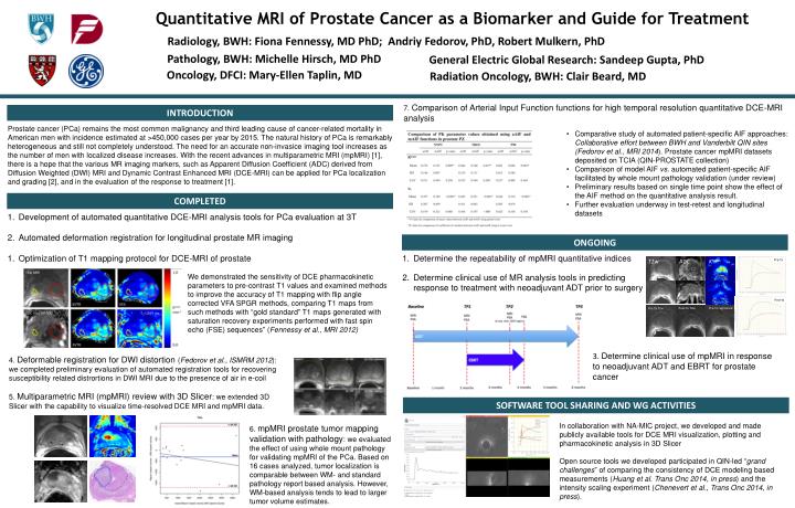
Quantitative MRI of Prostate Cancer as a Biomarker and Guide for Treatment. T2w. ADC. K trans. Pre-Tx. Radiology, BWH : Fiona Fennessy, MD PhD; Andriy Fedorov, PhD, Robert Mulkern, PhD. Pathology, BWH : Michelle Hirsch, MD PhD. General Electric Global Research : Sandeep Gupta, PhD.

E N D
Quantitative MRI of Prostate Cancer as a Biomarker and Guide for Treatment T2w ADC Ktrans Pre-Tx Radiology, BWH: Fiona Fennessy, MD PhD; Andriy Fedorov, PhD, Robert Mulkern, PhD Pathology, BWH: Michelle Hirsch, MD PhD General Electric Global Research: Sandeep Gupta, PhD Post-tx Post-Tx T2w Pre-Tx registered Pre-Tx T2w Oncology, DFCI: Mary-Ellen Taplin, MD Radiation Oncology, BWH: Clair Beard, MD 7. Comparison of Arterial Input Function functions for high temporal resolution quantitative DCE-MRI analysis INTRODUCTION Prostate cancer (PCa) remains the most common malignancy and third leading cause of cancer-related mortality in American men with incidence estimated at >450,000 cases per year by 2015. The natural history of PCa is remarkably heterogeneous and still not completely understood. The need for an accurate non-invasice imaging tool increases as the number of men with localized disease increases. With the recent advances in multiparametric MRI (mpMRI) [1], there is a hope that the various MR imaging markers, such as Apparent Diffusion Coefficient (ADC) derived from Diffusion Weighted (DWI) MRI and Dynamic Contrast Enhanced MRI (DCE-MRI) can be applied for PCa localization and grading [2], and in the evaluation of the response to treatment [1]. • Comparative study of automated patient-specific AIF approaches: Collaborative effort between BWH and Vanderbilt QIN sites (Fedorov et al., MRI 2014). Prostate cancer mpMRI datasets deposited on TCIA (QIN-PROSTATE collection) • Comparison of model AIF vs. automated patient-specific AIF facilitated by whole mount pathology validation (under review) • Preliminary results based on single time point show the effect of the AIF method on the quantitative analysis result. • Further evaluation underway in test-retest and longitudinal datasets COMPLETED • Development of automated quantitative DCE-MRI analysis tools for PCa evaluation at 3T • Automated deformation registration for longitudinal prostate MR imaging • Optimization of T1 mapping protocol for DCE-MRI of prostate ONGOING • Determine the repeatability of mpMRI quantitative indices • Determine clinical use of MR analysis tools in predicting response to treatment with neoadjuvant ADT prior to surgery We demonstrated the sensitivity of DCE pharmacokinetic parameters to pre-contrast T1 values and examined methods to improve the accuracy of T1 mapping with flip angle corrected VFA SPGR methods, comparing T1 maps from such methods with “gold standard” T1 maps generated with saturation recovery experiments performed with fast spin echo (FSE) sequences” (Fennessy et al., MRI 2012) 3. Determine clinical use of mpMRI in response to neoadjuvant ADT and EBRT for prostate cancer 4. Deformable registration for DWI distortion (Fedorov et al., ISMRM 2012): we completed preliminary evaluation of automated registration tools for recovering susceptibility related distrortions in DWI MRI due to the presence of air in e-coil 5. Multiparametric MRI (mpMRI) review with 3D Slicer: we extended 3D Slicer with the capability to visualize time-resolved DCE MRI and mpMRI data. SOFTWARE TOOL SHARING AND WG ACTIVITIES In collaboration with NA-MIC project, we developed and made publicly available tools for DCE MRI visualization, plotting and pharmacokinetic analysis in 3D Slicer Open source tools we developed participated in QIN-led “grand challenges” of comparing the consistency of DCE modeling based measurements (Huang et al. Trans Onc 2014, in press) and the intensity scaling experiment (Chenevert et al., Trans Onc 2014, in press). 6. mpMRI prostate tumor mapping validation with pathology: we evaluated the effect of using whole mount pathology for validating mpMRI of the PCa. Based on 16 cases analyzed, tumor localization is comparable between WM- and standard pathology report based analysis. However, WM-based analysis tends to lead to larger tumor volume estimates.