Download
1 / 27
310 likes | 1.04k Views
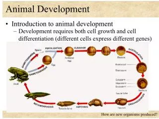
Animal Development. Chapter 47. Three Stages of Embryonic Development. Embryonic development is a succession of mitotic divisions which result in the single cell zygote becoming a complete organism consisting of trillions of cells. 1. Cell division: increase in the number of cells.

E N D
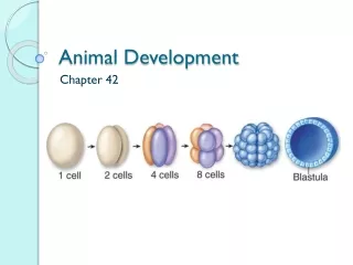
Animal Development Chapter 47
Three Stages of Embryonic Development • Embryonic development is a succession of mitotic divisions which result in the single cell zygote becoming a complete organism consisting of trillions of cells. • 1. Cell division: increase in the number of cells. • 2. Differentiation: development of specialized cells that are organized into tissues and organs. • 3. Morphogenesis: physical processes that give shape to an animal's body and organs. • Precise regulation of the three developmental processes is necessary for proper development. A breakdown in this regulation can result in developmental disorders. • • Cleft palate results when the upper palate fails to close. • • Webbed fingers and toes result from the failure of normal cell death.
Eggs: Bird, Reptiles, Amphibians • Largest yolk mass of animal groups. • Bird and reptile eggs have hard or leathery outer covering. • Area where yolk is most concentrated is vegetal pole. • Area where yolk is less concentrated is animal pole.
Eggs: Placental Mammals • Central yolk region surrounded by cytoplasm. • Yolk supplies nutrients for brief period of time until implantation in uterus. • Surrounded by jelly coat, follicle cells, and vitelline layer.
Fertilization… • …forms a diploid zygote from the haploid sets of chromosomes from two individuals, and triggers embryonic development. • Acrosomal reaction -- hydrolytic enzymes released from the acrosome of the sperm cell to digest jelly layer of egg. • Bindin (protein) coating on the acrosome attaches to receptors on the egg's vitelline layer; species specific. • Sperm and egg's plasma membranes fuse, allowing the sperm nucleus to enter the egg. • The membrane depolarizes (Na+ flows into the egg) to prevent other sperm cells from fusing (fast block to polyspermy).
Fertilization (cont) • Cortical Reaction -- release of calcium (Ca2+) from the egg cell's endoplasmic reticulum. • Increase in Ca causes cortical granules to fuse with the plasma membrane and release enzymes and polysaccharides into the perivitelline space (between cell membrane and vitelline layer). • Separation of the vitelline layer from the plasma membrane occurs. • Osmosis into the space causes the area to swell; vitelline layer hardens to form the fertilization membrane (slow block to polyspermy).
Egg Activation • Increased Ca concentration also stimulates cellular respiration and protein synthesis rates in the egg cell. • H+ is pumped out of the cell, changing pH changes from slightly acidic (6.8) to slightly alkaline (7.3). • Activation can be artificially induced by injection of Ca+; development will begin without fertilization. • Chromosomes from the sperm nucleus unite with the egg nucleus to form the zygote (actual fertilization).
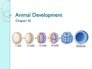
Cleavage • Cleavage is a succession of rapid mitotic cell divisions following fertilization that produces a multicellular embryo, the blastula. • During cleavage, the cells undergo the S and M phases of the cell cycle but the G1 and G2 phases are virtually skipped. • The zygote is divided into many smaller cells called blastomeres. • In frogs, the animal hemisphere has a gray hue due to the presence of melanin in the cytoplasm; the vegetal hemisphere has a light yellow hue due to the yolk. • Cleavage in the animal hemisphere of a frog's zygote is more rapid than in the vegetal hemisphere. • In sea urchins and many other animals, divisions to occur at about the same rate due to smaller amounts of yolk.
Cleavage (cont) • First two divisions in sea urchins and frogs are vertical; divide the embryo into four cells. • Third cleavage is horizontal and produces an eight cell embryo with two tiers (animal and vegetal) of four cells each. • • In deuterostomes (radial cleavage), the upper tier of cells is aligned directly over the lower tier. • • In protostomes (spiral cleavage), the upper tier of cells align with the grooves between cells of the lower tier. • The embryo becomes a solid ball of cells called a morula. • The blastocoel, a fluid-filled cavity, develops within the embryo which changes it to a hollow ball of cells, the blastula. • • In sea urchins, the blastocoel is centrally located in the blastula due to equal cell divisions. • • Unequal cell divisions in the frog embryo produces a blastocoel in the animal hemisphere.
Gastrulation (read pp. 592-594) • Gastrulation rearranges the blastula to form a three-layered embryo (gastrula) with a primitive gut • Three layers produced by gastrulation are tissues called embryonic germ layers. • Ectoderm (nervous system and outer layer of skin). • Endoderm (lining of the digestive tract and liver and pancreas). • Mesoderm (kidneys, heart, muscles, inner layer of the skin, and most other organs). • Animals are classified on the basis of how these tissues are organized. • Acoelomates – no body cavity between the gut and the body wall (flatworms). • Pseudocoelomates – have a body cavity, but not completely lined with mesoderm (roundworms). • Coelomates – have a “true coelom”, a body cavity completely lined with mesoderm; body cavities may develop differently. • Protostomes include mollusks, arthropods, and segmented worms. • Deuterostomes include echinoderms and vertebrates.
Gastrulation in sea urchins begins at the vegetal pole; single layer of cells in the blastula invaginates. Cells in this area detach and enter the blastocoel (cavity) as migratory mesenchyme cells. The area that is “collapsing” forms a pouch called the archenteron or primitive gut. The opening that is formed is the blastopore which will become the anus (deuterostomes) or the mouth (protostomes). Gastrulation during frog development begins when a small crease forms as a cluster of cells burrow inward (invagination). Involution then occurs as cells on the surface of the embryo roll over the fold into the embryo's interior. The circular blastopore surrounds a group of large, food-laden cells from the vegetal pole called the yolk plug. Gastrulation (cont)
http://www.gastrulation.org/Movie9_3.mov • http://www.luc.edu/faculty/wwasser/dev/devm.htm (Lots of good visuals!) • http://www.luc.edu/faculty/wwasser/dev/xlgast.mov (amphibian gastrulation external) • http://www.luc.edu/faculty/wwasser/dev/wholegas.mov (amphibian gastrulation internal)
http://courses.biology.utah.edu/gard/development/Images_and_movies/Gastrulation/Movies/dorsal_lip.movhttp://courses.biology.utah.edu/gard/development/Images_and_movies/Gastrulation/Movies/dorsal_lip.mov
Organogenesis • In humans, all major organs have developed from the three germ layers in the first 3 months of pregnancy; begins with neural tube formation in the gastrula. • Development of cells depends on: • 1. Location of the cells in the embryo -- cell fate depends on contents of cytoplasm which influence gene expression. • 2. Polarity of the embryo -- bilaterally symmetrical animals have an anterior-posterior axis, a dorsal-ventral axis, and left and right sides. • 3. Morphogenetic Movements -- cell extension, contraction, and adhesion guide movement and reorganization of cells. • 4. Induction -- ability of one cell group to influence the development of another.
Notochord: Primitive “spine”; induces neural tube to form above it; vertebral column forms around it; persists as your disks. • Neural tube : Becomes spinal cord and brain. • Neural crest: Cells migrate to form other nerves and bones. • Somites: Form vertebrae.
Organogenesis (cont) • 5. Differentiation -- cells become specialized as tissues and organs form in the embryo. • 6. Genomic equivalence – All cells have the same genes, but cells differ in structure and function because they express different portions of the genome. • 7. Pattern Formation -- body form with specialized organs and tissues all in their characteristic places; genes respond to positional info and chemical cues which vary with location. • Homeotic gene sequences are virtually identical in all animals; contains a “homeobox” sequence which produce a protein that activates or represses gene activity.