Download
1 / 5
50 likes | 53 Views
This tool allows you to manipulate the position of the virtual electrode to view different regions of the retina. Control the stimulus parameters and see the firing rates of the cells. Explore and record from different columns in the cortex.

E N D
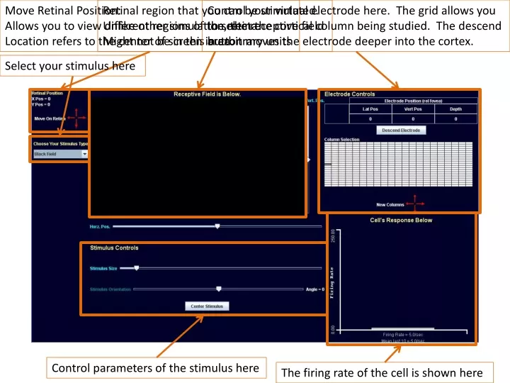
Move Retinal Position: Allows you to view different regions of the retina. Location refers to the center of screen in arbitrary units Retinal region that you can be stimulated. Unlike other simulation, the receptive field Might not be in this area. Control your virtual electrode here. The grid allows youto select the cortical column being studied. The descendbutton moves the electrode deeper into the cortex. Select your stimulus here Control parameters of the stimulus here The firing rate of the cell is shown here
Move Retinal Position: The red arrows will move in the indicated direction¼ the visible area in that direction. That is, if the center is 0 and the right edge is 50,one click on the right red arrow will put the centerat 25. It is the same proportion, not absolute movementfor the vertical arrows. Viewing Different Areas of the Retina The position of the center of the screen on the fovea isindicated here. The units are arbitrary. Think of them as mm.The position 0,0 indicates the fovea.
Select your stimulus here. Controlling the Stimulus You can move your stimulus across the visible retinal area by eitherdragging your mouse over the area or using the sliders along the sides. With these controlsyou can change thebar or dot width,the bar tilt (orientation)through -90 to +90 deg,and put the stimulusin the center of the visible retina. If the moving bar is used, youcan reverse the direction of motion. If present, there is acheckbox you can select to show the receptive field.
Output of the Cell Cell’s output. Think of the value as being number of actionpotentials a second.
Current Position of Electrode:Recall that the cortex is divided into vertical columns. The lateral and vertical position is the current column being studied relative to the first column. The first column is the left edge of the Striate cortex, centered vertically. This will be the fovea. You can only move to the right but both up and down. The depth is the distance from the surface in number of cells recorded from. Controlling the Electrode Pressing this button will allow you to recordfrom a cell deeper in the cortex. You can record from 25 cells in any column. Oncea cell has been left it cannot be accessed again Each white square represent a different column on the cortex. Click on that square to start recording from that cortex, starting witha cell nearest the surface. When you select acolumn, that column will be grayed out. Once you leave a column you cannot again record from that column again. This control will allow you to select a new region of brain whose column you wish torecord from. Initially you can move up, down, and to the right. Once you have lefta brain region, you cannot return and record from any other cells in that region.