Download
1 / 101
1.04k likes | 1.27k Views
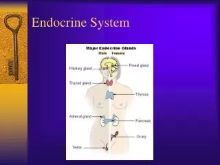
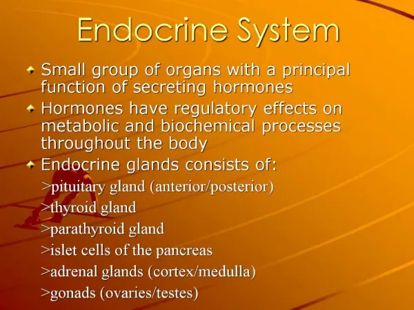
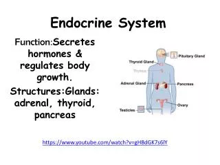
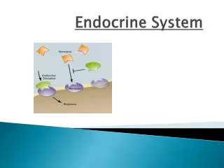
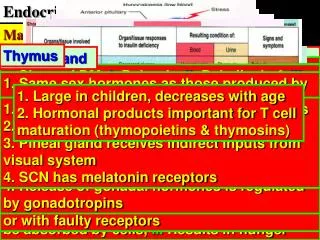
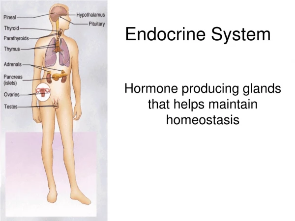
Endocrine System. It consists of two main components : The classical endocrine organs: pituitary, thyroid, parathyroid, adrenal, islets of langerhans in pancreas, ovary, testis and pineal gland.

E N D
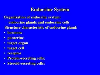
Endocrine System It consists of two main components: The classicalendocrine organs: pituitary, thyroid, parathyroid, adrenal, islets of langerhans in pancreas, ovary, testis and pineal gland. The diffuseendocrine system: consists of cells scattered as single or small group of cells in various non-endocrine organs such as lung, GIT, skin.
The pituitary gland: Wt. about 600 mg The pituitary gland consists of: anterior pituitary(adenohypophysis) and posterior pituitary (neurohypophysis). Anterior pituitary secretes growth hormone, prolactin, TSH, ACTH, FSH and LH . Posterior pituitary secretes oxytocin and ADH (vasopressin).
The normal microscopic appearance of the pituitary gland is shown here. The adenohypophysis is at the right and the neurohypophysis is at the left.
The normal microscopic appearance of the adenohypophysis is shown here. The adenohypophysis contains three major cell types: acidophils(40%), basophils(10%), and chromophobes(50%). The staining is variable, and to properly identify specific hormone secretion, immunohistochemical staining is necessary. A simplistic classification is as follows: • The pink acidophils secrete growth hormone (GH) and prolactin(PRL) • The dark purple basophils secrete corticotrophin (ACTH), thyroid stimulating hormone (TSH), and gonadotrophins: follicle stimulating hormone-luteinizing hormone (FSH and LH) • The pale staining chromophobes have few cytoplasmic granules, but may have secretory activity.
Pathology of adenohypophysis (Tumors): Pituitary adenoma: most common lesion of anterior pituitary, it divided into microadenoma (<15 mm in diameter) and macroadenoma (>15 mm in diameter). Effects or clinical features: • Endocrine effect due to hypersecretion of specific hormones. • Local pressure symptoms of large tumor causing compression on surrounding structure, optic chiasma,optic nerve, causing visual disturbance • pressure atrophy on adjacent normal gland lead to hypopituitarism. • Enlargement and erosion of the floor of the sellaturcica are common findings and important radiological signs.
Pituitary adenoma. The circumscribed mass lesion present here in the sella turcica is a pituitary adenoma. Though pituitary adenomas are benign, they can produce problems either from a mass effect (usually visual problems from pressing on the optic chiasm and/or headaches) or from production of hormones such as prolactin or ACTH.
This is a microadenoma of the anterior pituitary. Such microadenomas may appear in 1 to 5% of adults. These microadenomas rarely have a significant hormonal output that leads to clinical disease.
The microscopic appearance of the pituitary adenoma is shown here. Note the monotonous appearance of these small round cells with small round nuclei and pink to blue cytoplasm. The cells may be arranged in nests or cords and endocrine tumors also have prominent vascularity.
Other tumors • Pituitary carcinoma:extremely rare • Craniopharyngioma :Most patients with craniopharyngioma are in the first or second decade of life. • Its location is usually suprasellar, although it may occupy the sella as well • Metastatic tumors :from breast, lung ,GIT. Posterior lobe is mostly affected, anterior lobe rarely involved.
A craniopharyngioma is seen here at medium and high power. It is derived from remnants of Rathke's pouch and forms an expanding mass arising in the sella turcica that erodes bone and infiltrates into surrounding structures. They are difficult to eradicate, even though they are composed of histologically appearing squamoid and columnar epithelium lining cystic spaces filled with oily fluid.
Other pathology; • Inflammatory conditions • Circulatory disturbances (Sheehan's syndrome): necrosis of anterior lobe due to severe post-partum hemorrhage after labour and the patient develop after that hypopituitarism.
Hyperfunction of adenohypophysis • Hypersecretion of GH: Acromegaly and gigantism. • ACTH: Cushing’s disease. • Prolactin: hyperprolactinemia
Acromegaly and gigantism: • Cause: pituitary adenoma. • Gigantism: when hypersecretion of GHoccur in children before closure of epiphysis result in proportional increase in length and thickness of bones with delayed in epiphyseal fusion. If excess of GH persists after epiphyseal closure causing feature of acromegaly.
Acromegaly: occur when hyper-secretion of GH occur after fusion of epiphysis in adult. clinical features; enlargement of extremities, over-growth of bone, and soft tissue, large hand and feet, enlargement of nose and lower jaw (prognathism), cardiomegally, hypertension, D.M., osteoarthritis, kyphosis, increased sweating, 30-40% have hyper-prolactinaemia leading to galactorrhoea.
Cushing’s disease: hypersecretion of ACTH result in adrenal gland hyperplasia with excessive secretion of glucocorticoids by adrenalsresulting in Cushing’s syndrome .
Causes of ACTH hypersecretion: • ACTH-cell adenoma. • ACTH-cell hyperplasia. • Ectopic ACTH secretion
Hyperprolactinemia: Causes: • Adenoma (prolactinoma). • destructive lesion of hypothalamus. • drugs (methyldopa, phenothiazine). • physiological type in pregnancy.
In female it results in amenorrhea, infertility, galactorrhoea, • in men it is usually asymptomatic, may cause loss of libido, infertility, impotence.
Hypopituitarism: Causes: • Pituitary tumor with pressure atrophy • Sheehan’s syndrome in developing countries • Pituitary surgery or radiotherapy • Trauma • Inflammation (tuberculosis, sarcoidosis, syphilis) • Autoimmune disease • Histiocytosis X • Craniopharyngioma • Metastatic tumors
Neurohypophysis: • ADH: reabsorption of water by renal tubules and concentration of urine. • Its deficiency causes diabetes insipidus: polyuria, polydipsia with diluted urine which differentiated it from psychogenic polydipsia. Causes: head injury, surgical trauma, destructive lesion of hypothalamus (sarcoidosis, histiocytosis-X), tumors, idiopathic. • Excessive secretion of ADH cause inappropriate secretion of ADH, water retention causing hyponatraemia, hypoosmolarity resulting in vomiting, muscle cramps, weakness, central edema which may lead to coma, death. Causes: ectopic secretion of ADH by small cell CA. of the lung, pneumonia, head injury, meningitis, subarachnoid hemorrhage and idiopathic.
Hypothalamus: Tumors: • Gliomas and germinomas. • Gangliocytoma Histologically: this lesion is composed of a mixture of mature neurons, astrocytes, and oligodendrocytes arranged with a varying degree of organization.
Normal thyroid gland :This is the normal appearance of the thyroid gland on the anterior trachea of the neck. The thyroid gland has a right lobe and a left lobe connected by a narrow isthmus. The normal weight of the thyroid is 10 to 30 grams. It cannot easily be palpated on physical examination.
Normal thyroid seen microscopically consists of follicles lined by an epithelium and filled with colloid .This normal thyroid follicle is lined by a cuboidal follicular epithelium with cells that can add or subtract colloid depending upon the degree of stimulation from TSH (thyroid stimulating hormone) released by the pituitary gland. As in all endocrine glands, the interstitium has a rich vascular supply into which hormone is secreted.
Congenital abnormalities: • Congenital absence of thyroid (aplasia) causes cretinism. • Ectopic thyroid gland: lingual thyroid, along midline in neck: sublingual, suprahyoid, infrahyoid or lateral position in neck (lateral aberrant thyroid) which always represent lymph node metastasis from thyroid carcinoma (usually papillary carcinoma) rather than ectobia. Rare sites esophagus, larynx, trachea, soft tissues of the neck. • Thyroglossal fistula and thyroglossal cyst in midline of the neck lined by respiratory or squamous epithelium, contain thyroid tissue and lymphoid tissue in its wall.
Thyroid diseases present either as thyroid enlargement (goiter: diffuse or nodular) or as excess (Hyperthyroidism) or deficiency (hypothyroidism) of thyroid hormones. Hyperthyroidism: Clinically called thyrotoxicosis due to hypersecretion of thyroid hormones and presents with signs of hypermetabolism and excessive stimulation of the sympathetic system (patient present with weight loss, but increased sensitivity to circulatory adrenaline, and there is increased appetite,patient is nervous, irritable with heat intolerance, excessive sweating, fine tremor, tachycardia, atrial fibrillation, which may lead to cardiac failure, eye signs include; lid-lag and lid-retraction.
Causes: • Grave’s disease is the most common cause in 80-85% of cases. • Toxic nodular goiter 10% of cases. • Thyroid adenoma 5-10% of cases. • Other causes: Early Hashimoto’s thyroiditis, pituitary adenoma secrete TSH, exogenous thyroid hormone and large doses of iodine give to patient with nontoxic nodular goitre
Hypothyroidism: Deficiency of thyroid hormones in adult is called myxoedema and in infant and early childhood is called cretinism. Myxedema:dry waxy swelling of the skin of the extremities and face. patient is lethargic, feel cold, with constipation, psychosis, skin and hair are dry, coarse facial feature due to deposition of mucopolysaccharide in dermis, and there is pain, parasthesia (involvement of nerves), voice is gruff (involvement of larynx), increased weight, increased serum cholesterol, bradycardia, pericardial effusion . Cretinism: mental retardation, dwarfism, coarse facial features, a protruding tongue and umbilical hernia.
Causes of hypothyroidism: • Autoimmune thyroiditis such as Hashimoto’s thyroiditis (most common cause in adult) and primary myxoedema. • Congenital: aplasia or hypoplagia. • Iatrogenic: following thyroidectomy, radiation therapy. • Secondary hypothyroidism due to hypopituitarism. • Severe iodine deficiency (in endemic areas). • Dietary: goitrogenes in food • Drugs: propylthyouracil, lithium. • Genetic causes; include; dyshormogenetic goiter, due to absence of enzymes involved in synthesis of thyroid hormones. • Pendred’s syndrome (dyshormogenesis+deafness+mutism).
Goiter:is enlargement of the thyroid. It is either diffuse or nodular, nontoxic or toxic. Nontoxic goiter : Commonest lesion of thyroid gland, causing enlargement of thyroid as a compensatory hyperplasia by increase TSH due to defect in the synthesis of thyroid hormones. It is of two types: • Endemic goiter • Sporadic goiter
Endemic goiter: Occurrence of nontoxic goiter in more than 10% of population. It is always related to iodine deficiency. Usually occurs in mountainous areas and areas remote from the sea, it is due to iodine deficiency in food and water, but its incidence has decreased upon introduction of ionized salt. • Morphological features is the same for sporadic and endemic goiter.
Nodular goiter • This patient was euthyroid. This represents the most common cause for an enlarged thyroid gland and the most common disease of the thyroid. • MNG characterized by • Asymmetrical enlargement. • Multiple nodules of variable size. • Secondary degenerative changes: hemorrhage, fibrosis, cystic degeneration filled with fluid, and calcification can be formed.
Microscopic features of nontoxic goiter: • Early stage, diffuse hyperplasia of follicles with scanty colloid (parenchymatous goiter), this is followed by accumulation of colloid with involution of epithelium (colloid goiter). • Later on, nodules formation of variable size contain colloid separated by fibrous tissue (multinodular colloid goiter) associated with degenerative changes: cystic changes, hemorrhage, fibrosis, calcification.
Multinodular goiter: Different sized follicles some are dilated and lined by flattened epithelium (indicate inactivity), filled with colloid,others lined by normal or hyperplastic epith.
In some cases, one nodule prominently enlarged (dominant nodule) confused with tumor. In some cases of long standing multinodular goiter especially in elderly, show picture of hyperthyroidism (toxic nodular goiter).
Autoimmune thyroid diseases: These characterized by: • Presence of circulating auto antibodies to thyroid tissue. • Lymphocytic infiltration with destruction of thyroid tissue and formation of lymphoid follicles with germinal centers. • It may be associated with other autoimmune diseases such as Addison’s disease, D.M., SLE, rheumatoid arthritis, pernicious anemia. they include:
Hashimoto’s thyroiditis: • this disease due to presence of auto antibodies (antithyroglobulin, anti-microsomal antibody). • Increase in HLA-DR5 and B5 suggests genetic predisposition. • 1-2% led to B-cell lymphoma. • Increase risk of papillary carcinoma.
This symmetrically small thyroid gland demonstrates atrophy. This patient was hypothyroid. This is the end result of Hashimoto's thyroiditis. Initially, the thyroid is enlarged and there may be transient hyperthyroidism, followed by a euthyroid state and then hypothyroidism with eventual atrophy years later.
Here is a low power microscopic view of a thyroid with Hashimoto's thyroiditis. Note the lymphoid follicle at the right center. This is an autoimmune disease and often antithyroglobulin and antimicrosomal antibodies can be detected. Other autoimmune diseases such as Addison's disease or pernicious anemia may also be present.
Hashimoto’s thyroiditis: 1- Hürthle cells: large pink cells at the center and right 2- The lymphoid follicle with germinal center is at the left.
Grave’s disease: • It’s characterized by diffuse thyroid hyperplasia and hyperthyroidism (diffuse toxic goiter) • Its incidence increase in HLA-DR3 individuals suggests genetic predisposition. • It results from presence of autoantibodies to TSH receptors cause their activation and stimulate thyroid hormone secretion causing hyperthyroidism. These antibodies are called thyroid stimulating immunoglobulin (TSI: stimulate thyroxin -T4 synthesis). TGI lead to gland hyperplasia and enlargement .
Grave’s disease (difuse toxic goiter) A diffusely enlarged thyroid gland associated with hyperthyroidism is known as Grave's disease. Note the infoldings of the hyperplastic epithelium line by tall columnar thyroid epithelium with clear vacuoles in the colloid next to the epithelium where the increased activity of the epithelium to produce increased thyroid hormone has led to scalloping out of the colloid .
Other autoimmune diseases • Primary myxoedema. • Lymphocytic thyroiditis.
Other type of thyroiditis: • De Quervain’s thyroiditis(granulomatous, or subacute thyroiditis). • Riedel’s thyroiditis
DeQuervian’s thyroiditis:This is subacute granulomatous thyroiditis which probably follows a viral infection and leads to a painful enlarged thyroid. This disease is usually self-limited over weeks to months and the patients return to euthyroid state. Note the foreign body giant cells with destruction of thyroid follicles.
Riedel’s thyroiditis Very rare disease of unknown etiology, characterized by extensive replacement of thyroid tissue by dense fibrous tissue causing hardness of the gland (stony hard) with extension of fibrous tissue outside the gland cause fixation of thyroid to adjacent structures (iron collar) such as: trachea, recurrent laryngeal nerve (clinically mimic Carcinoma), some cases associated with retroperitoneal or mediastinal fibrosis
Thyroid tumors: 1. Tumors arise from follicular epithelial cells: • benign: follicular adenoma (majority of thyroid tumor) • malignant: follicular carcinoma, papillary carcinoma and anaplastic carcinoma. 2.Tumors arise from C cells: medullary carcinoma.
Follicular adenoma: • Commonest thyroid tumor mainly in females over 30 years. Adenoma usually presents with nonfunctioning (cold) nodule but may be cause hyperthyroidism (toxic adenoma) which is hot nodule on thyroid scan. The nodule is painless, if large may produce local symptoms. • Gross:Adenoma usually solitary encapsulated nodule (3-10 cm) and compressing surrounding thyroid tissue. • Histopathological features: Adenoma consists of uniform follicles contain colloid, surrounded by fibrous capsule without capsular or vascular invasion with or without nuclear pleomorphism or atypia (endocrine atypia). • Adenoma with microfollicles and little colloid called fetal adenoma, while those with macrofollicles filled with colloid called colloid adenoma. • If it consists of Hürthle cells (Hürthle cell adenoma). • All have the same behavior.
Follicular neoplasm ( follicular adenoma histologically) A solitaryneoplasm that is surrounded by a thin white capsule. It is sometimes difficult to tell a well-differentiated follicular carcinoma from a follicular adenoma. Thus, patients with follicular neoplasms are treated with subtotal thyroidectomy just to be on the safe side.