Download
1 / 42
430 likes | 456 Views
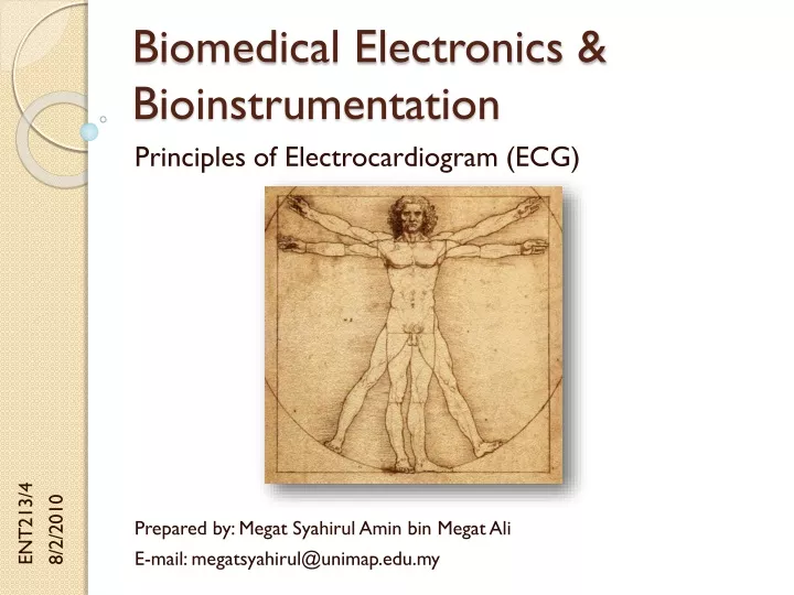
Biomedical Electronics & Bioinstrumentation. Principles of Electrocardiogram (ECG). ENT213/4 8/2/2010. Prepared by: Megat Syahirul Amin bin Megat Ali E-mail: megatsyahirul@unimap.edu.my. Contents. The Heart Revisited Standard Lead System The Einthoven Triangle

E N D
Biomedical Electronics & Bioinstrumentation Principles of Electrocardiogram (ECG) ENT213/4 8/2/2010 Prepared by: MegatSyahirulAmin bin Megat Ali E-mail: megatsyahirul@unimap.edu.my
Contents • The Heart Revisited • Standard Lead System • The Einthoven Triangle • Functional Blocks of ECG • The Wilson Network & Central • The ECG Preamplifier • Noise Reduction Techniques • Protection Schemes
The Heart Revisited • The heart is a muscle formed in a way that allows it to act as a pump for blood. • The heart pumps blood when the muscle cells making up the heart wall contract, generating their action potential. • This potential creates electrical currents that spread from the heart throughout the body.
The Heart Revisited • The spreading electrical currents create differences in electrical potential between various locations in the body, and these potential can be detected and recorded through surface electrodes attached to the skin. • The waveform produced by these biopotentials is called the electrocardiogram (ECG), that is a written record (graph) of the cardiac electrical potential waveform.
ECG Waveform • Typical ECG waveform is shown below. The particular waveform is typical of a measurement from right arm to left arm.
ECG Waveform • The figure below shows the voltage amplitude relationship. A 1mV calibration pulse is also shown.
Standard Lead System • There are five electrodes connected to the patient in standard ECG recording. • Right arm (RA) • Left arm (LA) • Left leg (LL) • Right leg (RL) • Chest (C) • These electrodes are connected to the inputs of a differential buffer amplifier through lead selector switch.
Standard Lead System • The recording obtained across different pairs of electrodes result in different waveform shapes and amplitudes. • These different views are called leads. • Each lead conveys a certain amount of unique information that is not available in the other leads. • The physician is often able to diagnose the type and site of heart disease by examining these different views because the waveform anomalies have been correlated with disease conditions in the past.
Standard Lead System • The ECG machine uses the patient’s right leg as the common electrode, and the lead selected switch connects the proper limb or chest electrodes to the differential amplifier input.
Electrical Axis of the Heart • The figure shows the electrical axis of the heart that is examined by Leads I, II, III, aVR, aVF and aVL.
Bipolar Limb Leads • The bipolar limb leads are those designated Lead I, Lead II and Lead III that forms the Einthoven Triangle. • Lead I: LA to the amplifier’s non-inverting input, while RA is to the inverting input. • Lead II: The LL to the amplifier’s non-inverting input, while the RA is to the inverting input (LA shorted to RL). • Lead III: The LL to the amplifier’s non-inverting input, while the RA is to the inverting input (LA shorted to RL).
Bipolar Limb Leads • The electrical connections for the standard Bipolar Limb Leads:
Unipolar Limb Leads • The unipolar limb leads, also known as the augmented limb leads, examine the composite potential from all three limbs simultaneously. • In all three augmented limb leads, the signals from two limbs are summed in a resistor network and then applied to the amplifiers inverting input, while the signal from the remaining limb electrode is applied to the non-inverting input.
Unipolar Limb Leads • Lead aVR: RA to the non-inverting input, while LA and LL are summed at the inverting input. • Lead aVL: LA to the non-inverting input, while RA and LL are summed at the inverting input. • Lead aVF: LL to the non-inverting input, while RA and LA are summed at the inverting input.
Unipolar Limb Leads • The electrical connections for the standard Unipolar Limb Leads:
Unipolar Chest Leads • The unipolar chest leads (V1 through V6) are measured with the signals from certain specified locations on the chest applied to the amplifier’s non-inverting input, while RA, LA and LL signals are summed in a resistor Wilson network at the amplifier’s inverting input (called “indifferent electrode”).
Unipolar Chest Leads • The electrical connections for the standard Unipolar Chest Leads:
Functional Blocks of the ECG • Protection Circuit This circuit includes protection devices so that the high voltages that may appear across the input to the ECG under certain conditions do not damage it. • Lead Selector Each electrode connected to the patient is attached to the lead selector of the ECG. The function of this block is to determine which electrodes are necessary for a particular lead and to connect them to the remainder of the circuit.
Functional Blocks of the ECG • Calibration Signal A 1mV calibration signal is momentarily introduced into the ECG for each channel that is recorded. • Preamplifier The input preamplifier stage carries out the initial amplification of the ECG. This stage should have a very high input impedance and CMRR. A typical preamplifier stage is the differential amplifier that consists of three op-amps. A gain control switch is often included as a part of this stage.
Functional Blocks of the ECG • Isolation Circuit The circuitry of this block contains a barrier to the passage of current from the power line (50Hz). • Driven Right Leg Circuit The circuit provides a reference point on the patient that normally is at ground potential. This connection is made to an electrode on the patient’s right leg. • Drive Amplifier Circuitry of this block amplifies the ECG to a level which it can appropriately record the signal on the recorder.
Functional Blocks of the ECG • Memory System Many modern ECGs store in memory as well as printing them out on a paper chart. The signal is first digitized by an ADC, and then samples from each lead are stored in memory. Patient information entered via the keyboard is also stored. • Microcomputer The microcomputer controls the overall operation of the ECG. The operator can select several modes of operation by invoking a particular program.
Functional Blocks of the ECG • Recorder-Printer This block provides a hard copy of the recorded ECG signal. It also prints out the patient identification, clinical information entered by the operator, and the results of the automatic analysis of the ECG.
The Wilson Network & Central • The Wilson Network A passive resistor array that is used in deriving the six basic leads (I, II, III, aVR, aVL and aVF). • The Wilson Central Represents ECG zero and common-mode interference. It is derived from patient electrodes RA, LA, and LL and is actually the average of these (sum divided by 3).
The ECG Preamplifier • An ECG preamplifier is a differential bioelectric amplifier. • The input circuitry consists of: • The high-impedance input of the bioelectric amplifier. • Lead selector switch. • 1mV calibration source. • Means for protecting the amplifier against high-voltage discharges from defibrillators used on the patient.
The ECG Preamplifier • The amplifier may be the bioelectric instrumentation amplifier, although, in all modern machines, the isolation amplifier designs will be used for patient safety. • The common-mode voltage in the ECG case is composed of: • dc electrode offset potential. • 50Hz ac-induced interference. • Hum interference is caused by magnetic and electric fields from power lines and transformers cutting across ECG electrodes and patients.
Noise Reduction Techniques • Instrumentation Amplifier (IA): • The CMRR of the commercially available IA is very high and cancels some of the noise. • The differential amplification nature of the IA removes it, because equal common-mode (CM) noise is present on each IA input. • The IA just subtracts equal noise voltages to give nearly zero while amplifying the difference in the unequal ECG signals present on its inputs.
Noise Reduction Techniques • Right-Leg (RL) Drive: • The common-mode voltage (CMV) is inverted by the right-leg amplifier and the resultant voltage is applied to the patient’s right leg. • Just several microamperes or less are driven into the patient to prevent internal cardiac shock. • The circuit acts in a feedback loop (patient and electronics) to drive the CM noise on the patient to a low level. • Since the RL drive voltage is the inversion of the CMV, the right-leg goes in opposite direction compared to CMV on patient leads.
Noise Reduction Techniques • Isolation Amplifier: • Usually an isolation amplifier would follow the IA. • The iso-amp’s isolation-mode rejection (IMR) also reduces the noise by establishing 1012Ω and 9pF between the patient and earth ground. • Isolation acts to attenuate noise that is trying to mix with the low level ECG input signal. • Other CM Noise Reduction Method: • Shield Driver • 50Hz Notch Filter
Noise Reduction Techniques • The standard -3dB frequency response of the amplifier used in making diagnostic grade recordings is 0.05 to 100Hz, while monitoring instruments have a response of a bout 0.05 to 45Hz (varies between manufacturers). • ECG preamps must be ac-coupled so that artifacts from the electrode offset potential are eliminated. • The low-frequency response of the amplifier must not extend down to dc, but since certain features of the ECG waveform have a very low-frequency components, the response is nearly dc.
Protection Schemes • The defibrillator is a high-voltage electrical heart stimulator used to resuscitate heart attack victims. • It is necessary to have an ECG monitor connected to the patient when using the defibrillator, so the ECG preamplifier input must be designed to withstand high voltages and high peak currents, even though normal ECG waveforms are on the order of millivolts. • The high-voltage (over a kilovolt) burst from the defibrillator will last 5 to 20 ms.
Protection Schemes • Two stages of defibrillation protection: • Series resistors and neon bulbs. • Series resistors and clamp diodes.
Protection Schemes • Preamplifiers must also be protected from electrosurgical unit (ESU) high voltages. • The ESU interference can range from hundreds of kilohertz to 100MHz and up to several kilovolts which can greatly disrupt ECG signal. • The interferences can be reduced by using multistage: • RC filters. • LC filters.
Further Reading… • Carr, J.J. (2000). Introduction to Biomedical Equipment Technology. 4th Ed. Prentice Hall. • Chapter 8 • Webster, J.G. (2009). Medical Instrumentation: Application and Design. 4th Ed., Wiley. • Chapter 4 & 6
The End… “Shoot for the moon. Even if you miss it you will land among the stars…"