Download
1 / 28
310 likes | 645 Views
Microstructural characterization of Unidentified Bright Objects in Neurofibromatosis type 1. Can DKI and T2 relaxometry refine our understanding of tissue microstructure in NF1 related T2 hyperintensities ?. Thibo Billiet E-poster nr. 3654. Tuesday 23 april at 13:30, Computer 54.

E N D
Microstructural characterization of Unidentified Bright Objects in Neurofibromatosis type 1 Can DKI and T2 relaxometry refine our understanding of tissue microstructure in NF1 related T2 hyperintensities? Thibo Billiet E-poster nr. 3654 Tuesday 23 april at 13:30, Computer 54
Microstructural characterization of Unidentified Bright Objects in Neurofibromatosis type 1 Thibo Billiet1, Louise Emsell1, Burkhard Maedler2, Felice D'Arco3, Sabine Deprez1, Judith Verhoeven1, Ellen Plasschaert4, Ronald Peeters1, Alexander Leemans5, Bea Van den Bergh6, Mathieu Vandenbulcke7, Eric Legius4, and Stefan Sunaert1 1 Translational MRI, Imaging and Pathology dpt., KU Leuven, Leuven, Belgium Radiology, University Hospitals Leuven, Leuven, Belgium Leuven research Institute for Neuroscience & Disease, Leuven, Belgium Medical Imaging Research Center, KU Leuven & UZ Leuven, Leuven, Belgium Division of Stereotaxy and MR-Based Operative Techniques, Department of Neurosurgery, Bonn University Hospital, Bonn, Germany Radiology, University Federico II of Naples, Naples, Italy Human Genetics, KU Leuven, Leuven, Belgium Image Sciences Institute, University Medical Center Utrecht,Utrecht, The Netherlands Psychology, KU Leuven, Leuven, Belgium Psychiatry, KU Leuven, Leuven, Belgium
What are UBO’s? Discussion Research question Conclusion MWI results Materials & methods DTI & DKI results
What are UBO’s? Discussion Research question Conclusion MWI results Materials & methods DTI & DKI results
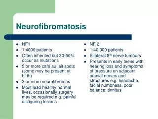
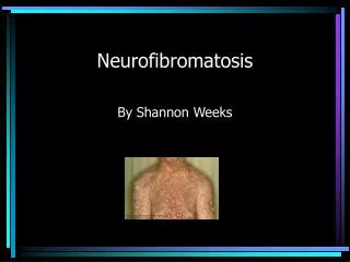
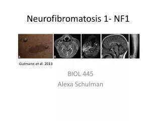
Unidentified Bright Objects are NF1-related T2 hyperintensities • Neurofibromatosis type 1: • Hereditary genetic disorder • Prevalence 1 in 3000 • Peripheral nerve tumours • Learning difficulties in children • … • (transient) hyperintensities on T2-weighted MRI scans Cerebellar white matter Basal ganglia and hypothalami Mesencephalon
Unidentified Bright Objects can be transient UBO number vs. age UBO volume vs. age Cerebellar WM Cerebellar WM Globus Pallidus / Internal Capsule Globus Pallidus / Internal Capsule Kraut M.A., Gerring J.P. et al., A.J.M.G. 129A:113-119 (2004)
The histopathological basis of UBOs remains unclear 1H MR Spectroscopy NAA: UBO < contralateral NAWM Choline: UBO > contralateral NAWM Jones, Gunawardena et al. 2001 Apparent Diffusion Coefficient (ADC) UBO > contralateral NAWM Tognini, Ferrozzi et al. 2005 Alkan, Sigirci et al. 2005 UBO > healthy control WM Eastwood, Fiorella et al. 2001 Van Engelen, Krab et al. 2008 Fractional Anisotropy (FA) UBO < healthy control WM Zamboni, Loenneker et al. 2007 Ferraz-Filho, da Rocha et al. 2011 Filippi, Bos et al. 2012 Magnetization Transfer Ratio (MTR) UBO < healthy control WM Margariti et al. 2007 Hamartomas? Braffman, Bilaniuk et al. 1988 Heterotopias? Bognanno, Edwards et al. 1988 Regions of abnormal myelination/demyelination? Smirniotopoulos and Murphy 1992 Intramyelinic vacuolization/astroglial cell proliferation? DiPaolo et al. 1995
The histopathological basis of UBOs remains unclear 1H MR Spectroscopy NAA: UBO < contralateral NAWM Choline: UBO > contralateral NAWM Jones, Gunawardena et al. 2001 Apparent Diffusion Coefficient (ADC) UBO > contralateral NAWM Tognini, Ferrozzi et al. 2005 Alkan, Sigirci et al. 2005 UBO > healthy control WM Eastwood, Fiorella et al. 2001 Van Engelen, Krab et al. 2008 Fractional Anisotropy (FA) UBO < healthy control WM Zamboni, Loenneker et al. 2007 Ferraz-Filho, da Rocha et al. 2011 Filippi, Bos et al. 2012 Magnetization Transfer Ratio (MTR) UBO < healthy control WM Margariti et al. 2007 Hamartomas? Braffman, Bilaniuk et al. 1988 Heterotopias? Bognanno, Edwards et al. 1988 Regions of abnormal myelination/demyelination? Smirniotopoulos and Murphy 1992 Intramyelinic vacuolization/astroglial cell proliferation? DiPaolo et al. 1995 Current hypothesis
What are UBO’s? Discussion Research question Conclusion MWI results Materials & methods DTI & DKI results
Can novel MRI techniques refine our understanding of UBO microstructure? DTI and DKI UBO vs. cNAWM Pairwise comparison ? MWI
What are UBO’s? Discussion Research question Conclusion MWI results Materials & methods DTI & DKI results
Excess Kurtosis = 0 Diffusion Tensor Imaging & Diffusion Kurtosis Imaging Radial diffusion No boundaries: Free diffusion Mean diffusion Fractional Anisotropy Axial diffusion Diffusion Tensor Imaging Excess Kurtosis = 0.5 Excess Kurtosis = 5 Kurtosis Tensor Imaging Axial kurtosis Kurtosis Anisotropy Mean kurtosis Radial kurtosis
Myelin Water Imaging A: the fraction of water with T2 relaxation time 10-40 ms correlates with the myelin content. This is the Myelin Water Fraction (MWF) B: The water having medium T2 (40-200 ms) is the intra- and extracellular water fraction (IEF). Note: The T2 time of CSF is even longer and not shown in this graph. C: the sum of all T2 fractions gives the total water content (TWC). D: the geometric mean T2 of the MWF peak (M-gmT2) E: the geometric mean T2 of total water content (T-gmT2) F: the geometric mean T2 of the IEF peak (IE-gmT2) G: the peak width of the IEF peak (IE-pw)
Data acquisition • MWI sequence • 3D GraSE • 32 echoes (TE1 = 10 ms, ΔTE = 10 ms) • TR = 800 ms • 32 slices (thickness 1mm) • FOV= 240 x 240 x 64mm3 • data matrix= 240 x 240 x 32 • EPI read-out factor = 3 • Madler and MacKay 2007 • Prasloski, Rauscher et al. 2012 • T2w-FLAIR • TR = 11000 ms • TI = 2800 ms • TE = 120 ms • Slice thickness 4 mm • FOV 230 x 184 x 119 mm3 • Data matrix 240 x 138 x 16 • DKI sequence • SE-EPI • 3 b0-images • B-shells: • 700 s/mm2 x 25 directions • 1000 s/mm2 x 40 directions • 2800 s/mm2 x 75 directions • δ/ Δ= 20ms/48.3ms • TR/TE= 7800ms/90ms • uniform voxel size= 2.5 mm • FOV= 240 x 240 x 125 mm3 • data matrix= 96 x 96 x 50 • SENSE factor = 2 in the anteroposterior direction • Poot, den Dekker et al. 2010 • Subjects • 7 NF1 patients (4 girls, 3 boys, mean age 12.6 years, SD 3.4 years) • 21 UBO-cNAWM pairs (DKI) / 10 UBO-cNAWM pairs (MWI)
What are UBO’s? Discussion Research question Conclusion MWI results Materials & methods DTI & DKI results
DTI results RD MD FA RD MD FA AD -- Increased diffusivity in radial direction is the main contributor to decreased FA and increased MD in UBOs No changes in axial direction
DKI results UBO radial compartmentalization RK MK KA cNAWM radial compartmentalization RK MK KA AK -- Decreased kurtosis in radial direction is the main contributor to decreased KA and decreased MK in UBOs No changes in axial direction
What are UBO’s? Discussion Research question Conclusion MWI results Materials & methods DTI & DKI results
MWI results UBO cNAWM In UBOs: 1) Longer overall T2 relaxation time
MWI results UBO cNAWM In UBOs: 1) Longer overall T2 relaxation time 2) Longer intra-and extracellular water T2
MWI results UBO cNAWM In UBOs: 1) Longer overall T2 relaxation time 2) Longer intra-and extracellular water T2 3) Extended range of T2 times in intra-and extracellular water
MWI results UBO cNAWM In UBOs: 1) Longer overall T2 relaxation time 2) Longer intra-and extracellular water T2 3) Extended range of T2 times in intra-and extracellular water 4) No changes in myelin water fraction (MWF) or intra-and extracellular water fraction (IEWF)
What are UBO’s? Discussion Research question Conclusion MWI results Materials & methods DTI & DKI results
What can MWI teach us about the T2 hyperintensities(UBOs)? • T2 hyperintensities arise in the intra- and extracellular space • Longer T2 relaxation times • Extended range of T2 times • Edema? • Barnes et al. 1987 (T2 and cerebral edema), Margariti et al. 2007 (MTR in UBOs), • Vacuolization? • Laule, Vavasour et al. 2007 (T2 and MS lesions) • Astroglial cell proliferation? • DiPaoloet al. 1995 (diffuse proliferation of protoplasmic astroglia in UBOs) • Unaltered MWF • No demyelination? • DiPaolo et al. 1995 (no effect on myelin stain in UBOs) UBO cNAWM
What is the added value of DTI & DKI? MWI results: T2 hyperintensities arise in the intra- and extracellular space UBO cNAWM Axial direction: no changes intact axons? DiPaolo et al. 1995 (no axonal damage in UBOs) Radial direction: Increased diffusivity + decreased kurtosis Intramyelinic vacuolization? DiPaolo et al. 1995 (spongiotic myelin in UBOs)
What are UBO’s? Discussion Research question Conclusion MWI results Materials & methods DTI & DKI results
Conclusion DTI and DKI UBO vs. cNAWM Pairwise comparison cNAWM MWI UBO • DiPaolo’s hypothesis confirmed: • No demyelination • No axonal damage • Intramyelinic vacuolization • Astroglial cell proliferation
Thank you for your attention Translational MRI – Advanced Neuroradiology Stefan Sunaert Ronald Peeters Sabine Deprez MarjoleinVerly Silvia Kovacs Sofie Van Cauter Louise Emsell