Download
1 / 61
620 likes | 877 Views
The Urinary System. Muse 2440 lecture #7 3/14/12. Overview of kidney functions. i. Regulation of blood ionic composition Regulation of blood pH Regulation of blood volume Regulation of blood pressure (hormone: Renin) Maintenance of blood osmolarity

E N D
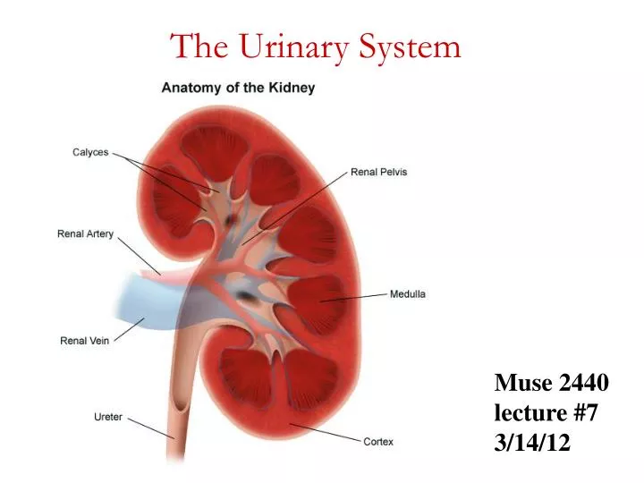
The Urinary System Muse 2440 lecture #7 3/14/12
Overview of kidney functions i • Regulation of blood ionic composition • Regulation of blood pH • Regulation of blood volume • Regulation of blood pressure (hormone: Renin) • Maintenance of blood osmolarity • Production of hormones (calcitrol and erythropoitin) • Regulation of blood glucose level • Excretion of wastes from metabolic reactions and foreign substances (drugs or toxins)
Anatomy and histology of the kidneys • External anatomy • Renal hilium – indent where ureter emerges along with blood vessels, lymphatic vessels and nerves • 3 layers of tissue • Renal capsule – deep layer – continuous with outer coat of ureter, barrier against trauma, maintains kidney shape • Adipose capsule – mass of fatty tissue that protects kidney from trauma and holds it in place • Renal fascia – superficial layer – thin layer of connective tissue that anchors kidney to surrounding structures and abdominal wall
Internal anatomy i • Renal cortex – superficial • Outer cortical zone • Inner juxtamedullary zone • Renal columns – portions of cortex that extend between renal pyramids • Renal medulla – inner region • Several cone shaped renal pyramids – base faces cortex and renal papilla points toward hilium • Renal lobe – renal pyramid, overlying cortex area, and ½ of each adjacent renal column
Anatomy of the kidneys • Parenchyma (functional portion) of kidney • Renal cortex and renal pyramids of medulla • Nephron – microscopic functional units of kidney • Urine formed by nephron drains into • Papillary ducts • Minor and major calyces • Renal pelvis • Ureter • Urinary bladder
Blood and nerve supply of the kidneys i • Blood supply • Although kidneys constitute less than 0.5% of total body mass, they receive 20-25% of resting cardiac output • Left and right renal artery enters kidney • Branches into segmental, interlobar, arcuate, interlobular arteries • Each nephron receives one afferent arteriole • Divides into glomerulus – capillary ball • Reunite to form efferent arteriole (unique) • Divide to form peritubular capillaries or some have vasa recta • Peritubular venule, interlobar vein and renal vein exits kidney • Renal nerves are part of the sympathetic autonomic nervous system • Most are vasomotor nerves regulating blood flow
Cortical radiate vein Cortical radiate artery Arcuate vein Arcuate artery Interlobar vein Interlobar artery Segmental arteries Renal vein Renal artery Renal pelvis Ureter Renal medulla Renal cortex (a) Frontal section illustrating major blood vessels Figure 25.4a
Aorta Inferior vena cava Renal artery Renal vein Segmental artery Interlobar vein Interlobar artery Arcuate vein Cortical radiate vein Arcuate artery Peritubular capillaries and vasa recta Cortical radiate artery Afferent arteriole Efferent arteriole Glomerulus (capillaries) Nephron-associated blood vessels (b) Path of blood flow through renal blood vessels Figure 25.4b
The nephron – functional units of kidney i • 2 parts • Renal corpuscle – filters blood plasma • Glomerulus – capillary network • Glomerular (Bowman’s) capsule – double-walled cup surrounding glomerulus • Renal tubule – filtered fluid passes into • Proximal convoluted tubule • Descending and ascending loop of Henle (nephron loop) • Distal convoluted tubule
Nephrons • Renal corpuscle and both convoluted tubules in cortex, loop of Henle extend into medulla • Distal convoluted tubule of several nephrons empty into single collecting duct • Cortical nephrons – 80-85% of nephrons • Renal corpuscle in outer portion of cortex and short loops of Henle extend only into outer region of medulla • Juxtamedullary nephrons – other 25-20% • Renal corpuscle deep in cortex and long loops of Henle extend deep into medulla • Receive blood from peritubular capillaries and vasa recta • Ascending limb has thick and thin regions • Enable kidney to secrete very dilute or very concentrated urine
Histology of nephron and collecting duct • Glomerular capsule • Visceral layer has podocytes that wrap projections around single layer of endothelial cells of glomerular capillaries and form inner wall of capsule • Parietal layer forms outer wall of capsule • Fluid filtered from glomerular capillaries enters capsular (Bowman’s) space
Basement membrane Podocyte Fenestrated endothelium of the glomerulus Glomerular capsule: visceral layer Figure 25.5
Renal tubule and collecting duct • Proximal convoluted tubule cells have microvilli with brush border – increases surface area • Juxtaglomerular appraratus helps regulate blood pressure in kidney • Macula densa – cells in final part of ascending loop of Henle • Juxtaglomerular cells – cells of afferent and efferent arterioles contain modified smooth muscle fibers • Last part of distal convoluted tubule and collecting duct • Principal cells – receptors for antidiuretic hormone (ADH) and aldosterone • Intercalated cells – role in blood pH homeostasis
Overview of renal physiology i • Glomerular filtration • Water and most solutes in blood plasma move across the wall of the glomerular capillaries into glomerular capsule and then renal tubule • Tubular reabsorption • As filtered fluid moves along tubule and through collecting duct, about 99% of water and many useful solutes reabsorbed – returned to blood • Tubular secretion • As filtered fluid moves along tubule and through collecting duct, other material secreted into fluid such as wastes, drugs, and excess ions – removes substances from blood • Solutes in the fluid that drains into the renal pelvis remain in the fluid and are excreted • Excretion of any solute = glomerular filtration + secretion - reabsorption
Renal tubule and collecting duct Renal tubule and collecting duct Renal tubule and collecting duct Renal corpuscle Renal corpuscle Renal corpuscle Afferent arteriole Afferent arteriole Afferent arteriole Glomerular capsule Glomerular capsule Glomerular capsule Urine (contains excreted substances) Urine (contains excreted substances) Urine (contains excreted substances) Fluid in renal tubule Fluid in renal tubule Fluid in renal tubule 1 1 1 Filtration from blood plasma into nephron Filtration from blood plasma into nephron Filtration from blood plasma into nephron 2 2 3 Tubular reabsorption from fluid into blood Tubular reabsorption from fluid into blood Tubular secretion from blood into fluid Efferent arteriole Efferent arteriole Efferent arteriole Blood (contains reabsorbed substances) Blood (contains reabsorbed substances) Blood (contains reabsorbed substances) Peritubular capillaries Peritubular capillaries Peritubular capillaries Structures and functions of a nephronsimplified schematic i
Glomerular filtration • Glomerular filtrate – fluid that enters capsular space • Daily volume 150-180 liters – more than 99% returned to blood plasma via tubular reabsorption • Filtration membrane – endothelial cells of glomerular capillaries and podocytes encircling capillaries • Permits filtration of water and small solutes • Prevents filtration of most plasma proteins, blood cells and platelets • 3 barriers to cross – glomerular endothelial cells fenestrations, basal lamina between endothelium and podocytes and pedicels of podocytes create filtration slits • Volume of fluid filtered is large because of large surface area, thin and porous membrane, and high glomerular capillary blood pressure
Podocyte of visceral layer of glomerular (Bowman’s) capsule Podocyte of visceral layer of glomerular (Bowman’s) capsule Podocyte of visceral layer of glomerular (Bowman’s) capsule Filtration slit Filtration slit Filtration slit Pedicel Pedicel Pedicel 1 1 1 Fenestration (pore) of glomerular endothelial cell: prevents filtration of blood cells but allows all components of blood plasma to pass through Fenestration (pore) of glomerular endothelial cell: prevents filtration of blood cells but allows all components of blood plasma to pass through Fenestration (pore) of glomerular endothelial cell: prevents filtration of blood cells but allows all components of blood plasma to pass through 2 2 Basal lamina of glomerulus: prevents filtration of larger proteins Basal lamina of glomerulus: prevents filtration of larger proteins 3 Slit membrane between pedicels: prevents filtration of medium-sized proteins (a) Details of filtration membrane (a) Details of filtration membrane (a) Details of filtration membrane Pedicel of podocyte Pedicel of podocyte Pedicel of podocyte Filtration slit Filtration slit Filtration slit Basal lamina Basal lamina Basal lamina Lumen of glomerulus Lumen of glomerulus Lumen of glomerulus 78,000x 78,000x 78,000x TEM TEM TEM Fenestration (pore) of glomerular endothelial cell Fenestration (pore) of glomerular endothelial cell Fenestration (pore) of glomerular endothelial cell (b) Filtration membrane (b) Filtration membrane (b) Filtration membrane
Net filtration pressure • Net filtration pressure (NFP) is the total pressure that promotes filtration • NFP = GBHP – CHP – BCOP • Glomerular blood hydrostatic pressure is the blood pressure of the glomerular capillaries forcing water and solutes through filtration slits • Capsular hydrostatic pressure is the hydrostatic pressure exerted against the filtration membrane by fluid already in the capsular space and represents “back pressure” • Blood colloid osmotic pressure due to presence of proteins in blood plasma and also opposes filtration
1 1 1 GLOMERULAR BLOOD HYDROSTATIC PRESSURE (GBHP) = 55 mmHg GLOMERULAR BLOOD HYDROSTATIC PRESSURE (GBHP) = 55 mmHg GLOMERULAR BLOOD HYDROSTATIC PRESSURE (GBHP) = 55 mmHg 2 2 CAPSULAR HYDROSTATIC PRESSURE (CHP) = 15 mmHg CAPSULAR HYDROSTATIC PRESSURE (CHP) = 15 mmHg 3 BLOOD COLLOID OSMOTIC PRESSURE (BCOP) = 30 mmHg Afferent arteriole Afferent arteriole Afferent arteriole Proximal convoluted tubule Proximal convoluted tubule Proximal convoluted tubule Efferent arteriole Efferent arteriole Efferent arteriole NET FILTRATION PRESSURE (NFP) =GBHP – CHP – BCOP = 55 mmHg 15 mmHg 30 mmHg = 10 mmHg NET FILTRATION PRESSURE (NFP) =GBHP – CHP – BCOP = 55 mmHg 15 mmHg 30 mmHg = 10 mmHg NET FILTRATION PRESSURE (NFP) =GBHP – CHP – BCOP = 55 mmHg 15 mmHg 30 mmHg = 10 mmHg Glomerular (Bowman's) capsule Glomerular (Bowman's) capsule Glomerular (Bowman's) capsule Capsular space Capsular space Capsular space
Glomerular filtration • Glomerular filtration rate GFR – amount of filtrate formed in all the renal corpuscles of both kidneys each minute • Homeostasis requires kidneys maintain a relatively constant GFR • Too high – substances pass too quickly and are not reabsorbed • Too low – nearly all reabsorbed and some waste products not adequately excreted • GFR directly related to pressures that determine net filtration pressure
3 Mechanisms regulating GFR • Renal autoregulation • Kidneys themselves maintain constant renal blood flow and GFR using • Myogenic mechanism – occurs when stretching triggers contraction of smooth muscle cells in afferent arterioles – reduces GFR • Tubuloglomerular mechanism – macula densa provides feedback to glomerulus, inhibits release of NO causing afferent arterioles to constrict and decreasing GFR
Mechanisms regulating GFR i • Neural regulation • Kidney blood vessels supplied by sympathetic ANS fibers that release norepinephrine causing vasoconstriction • Moderate stimulation – both afferent and efferent arterioles constrict to same degree and GFR decreases • Greater stimulation constricts afferent arterioles more and GFR drops • Hormonal regulation • Angiotensin II reduces GFR – potent vasoconstrictor of both afferent and efferent arterioles • Atrial natriuretic peptide increases GFR – stretching of atria causes release, increases capillary surface area for filtration
Tubular reabsorption and tubular secretion i • Reabsorption – return of most of the filtered water and many solutes to the bloodstream • About 99% of filtered water reabsorbed • Proximal convoluted tubule cells make largest contribution • Both active and passive processes • Secretion – transfer of material from blood into tubular fluid • Helps control blood pH • Helps eliminate substances from the body
Reabsorption routes and transport mechanisms • Reabsorption routes • Paracellular reabsorption • Between adjacent tubule cells • Tight junction do not completely seal off interstitial fluid from tubule fluid • Passive • Transcellular reabsorption – through an individual cell • Transport mechanisms • Reabsorption of Na+ especially important • Primary active transport • Sodium-potassium pumps in basolateral membrane only • Secondary active transport • Symporters, antiporters • Transport maximum (Tm) • Upper limit to how fast it can work • Obligatory vs. facultative water reabsorption
Reabsorption routes: paracellular reabsorption and transcellular reabsorption i
Reabsorption and secretion in proximal convoluted tubule (PCT) i • Largest amount of solute and water reabsorption • Secretes variable amounts of H+, NH4+ and urea • Most solute reabsorption involves Na+ • Symporters for glucose, amino acids, lactic acid, water-soluble vitamins, phosphate and sulfate • Na+ / H+antiporter causes Na+ to be reabsorbed and H+ to be secreted • Solute reabsorption promotes osmosis – creates osmotic gradient • Aquaporin-1 in cells lining PCT and descending limb of loop of Henle • As water leaves tubular fluid, solute concentration increases • Urea and ammonia in blood are filtered at glomerulus and secreted by proximal convoluted tubule cells
Reabsorption and secretion in the proximal convoluted tubule i
Microvilli Mitochondria Highly infolded plasma membrane Proximal convoluted tubule cells Figure 25.5
Reabsorption in the loop of Henle i • Chemical composition of tubular fluid quite different from filtrate • Glucose, amino acids and other nutrients reabsorbed • Osmolarity still close to that of blood • Reabsorption of water and solutes balanced • For the first time reabsorption of water is NOT automatically coupled to reabsorption of solutes • Independent regulation of both volume and osmolarity of body fluids • Na+-K+-2Cl- symporters function in Na+ and Cl- reabsorption – promotes reabsorption of cations • Little or no water is reabsorbed in ascending limb – osmolarity decreases
Na+–K+-2Cl- symporter in the thick ascending limb of the loop of Henle i
Reabsorption and secretion in the late distale convoluted tubule and collecting duct • Reabsorption on the early distal convoluted tubule • Na+-Cl- symporters reabsorb Na+ and Cl- • Major site where parathyroid hormone stimulates reabsorption of Ca+ depending on body’s needs • Reabsorption and secretion in the late distal convoluted tubule and collecting duct • 90-95% of filtered solutes and fluid have been returned by now • Principal cells reabsorb Na+ and secrete K+ • Intercalated cells reabsorb K+ and HCO3- and secrete H+ • Amount of water reabsorption and solute reabsorption and secretion depends on body’s needs
Hormonal regulation of tubular reabsorption and secretion i • Angiotensin II - when blood volume and blood pressure decrease • Decreases GFR, enhances reabsorption of Na+, Cl- and water in Proximal Convoluted Tubule • Aldosterone - when blood volume and blood pressure decrease • Stimulates principal cells in collecting duct to reabsorb more Na+ and Cl- and secrete more K+ • Parathyroid hormone • Stimulates cells in Distal Convolute Tubule to reabsorb more Ca2+
Antidiuretic hormone(ADH or vasopressin) Increases water permeability of cells by inserting aquaporin-2 in last part of DCT and collecting duct Atrial natriuretic peptide (ANP) Large increase in blood volume promotes release of ANP Decreases blood volume and pressure by inhibiting reabsorption of Na+ and water in PCT and collecting duct, suppress secretion of ADH and aldosterone Regulation of facultative water reabsorption by ADH
Production of dilute and concentrated urine i • Even though your fluid intake can be highly variable, total fluid volume in your body remains stable • Depends in large part on the kidneys to regulate the rate of water loss in urine • ADH controls whether dilute or concentrated urine is formed • Absent or low ADH = dilute urine • Higher levels = more concentrated urine through increased water reabsorption
Formation of dilute urine • Glomerular filtrate has same osmolarity as blood 300 mOsm/liter • Fluid leaving PCT is isotonic to plasma • When dilute urine is being formed, the osmolarity of fluid increases (concentrates) as it goes down the descending loop of Henle, decreases as it goes up the ascending limb, and decreases still more as it flows through the rest of the nephron and collecting duct
Osmolarity of interstitial fluid of renal medulla becomes greater, more water is reabsorbed from tubular fluid so fluid become more concentrated Water cannot leave in thick portion of ascending limb but solutes leave making fluid more dilute than blood plasma Additional solutes but not much water leaves in DCT Low ADH makes late DCT and collecting duct have low water permeability Formation of dilute urine i
Formation of concentrated urine • Urine can be up to 4 times more concentrated than blood plasma • Ability of ADH depends on presence of osmotic gradient in interstitial fluid of renal medulla • 3 major solutes contribute – Na+, Cl-, and urea • 2 main factors build and maintain gradient • Differences in solute and water permeability in different sections of loop of Henle and collecting ducts • Countercurrent flow of fluid though descending and ascending loop of Henle and blood through ascending and descending limbs of vasa recta
Countercurrent multiplication i • Process by which a progressively increasing osmotic gradient is formed as a result of countercurrent flow • Long loops of Henle of juxtamedullary nephrons function as countercurrent multiplier • Symporters in thick ascending limb of loop of Henle cause buildup of Na+ and Cl- in renal medulla, cells impermeable to water • Countercurrent flow establishes gradient as reabsorbed Na+ and Cl- become increasingly concentrated • Cells in collecting duct reabsorb more water and urea • Urea recycling causes a buildup of urea in the renal medulla • Long loop of Henle establishes gradient by countercurrent multiplication • organisms that adapt to deserts have long loops of Henle
Countercurrent exchange • Process by which solutes and water are passively exchanged between blood of the vasa recta and interstitial fluid of the renal medulla as a result of countercurrent flow • Vasa recta is a countercurrent exchanger • Osmolarity of blood leaving vasa recta is only slightly higher than blood entering • Provides oxygen and nutrients to medulla without washing out or diminishing gradient • Vasa recta maintains gradient by countercurrent exchange
Mechanism of urine concentration in long-loop juxtamedullary nephrons
Vasa recta Vasa recta Loop of Henle Loop of Henle Juxtamedullary nephron and its blood supply together Juxtamedullary nephron and its blood supply together Glomerular (Bowman’s) capsule Glomerular (Bowman’s) capsule H2O H2O Glomerulus Glomerulus Na+CI– Na+CI– Afferent arteriole Afferent arteriole Blood flow Blood flow Distal convoluted tubule Distal convoluted tubule Presense of Na+-K+-2CI– symporters Presense of Na+-K+-2CI– symporters Interstitial fluid in renal cortex Interstitial fluid in renal cortex Flow of tubular fluid Flow of tubular fluid 200 200 H H O O Efferent arteriole Efferent arteriole 2 2 H H O O 2 2 300 300 320 320 300 300 H H O O 2 2 Collecting duct Collecting duct Proximal convoluted tubule Proximal convoluted tubule 300 300 300 300 300 300 100 100 320 320 H2O H2O H2O H2O Na+CI– Na+CI– Na+CI– Na+CI– H2O H2O Interstitial fluid in renal medulla Interstitial fluid in renal medulla 400 400 380 380 200 200 400 400 400 400 500 500 H2O H2O 600 600 H2O H2O H2O H2O 580 580 400 400 600 600 Na+CI– Na+CI– 600 600 1 1 Osmotic gradient Osmotic gradient Symporters in thick ascending limb cause buildup of Na+ and Cl– Symporters in thick ascending limb cause buildup of Na+ and Cl– 700 700 H2O H2O 800 800 780 780 600 600 800 800 H2O H2O 800 800 Urea Urea H2O H2O 900 900 Na+CI– Na+CI– H2O H2O 1000 1000 980 980 800 800 1000 1000 1000 1000 1100 1100 H2O H2O Papillary duct Papillary duct 1200 1200 1200 1200 1200 1200 2 1200 1200 Countercurrent flow through loop of Henle establishes an osmotic gradient Loop of Henle Loop of Henle 1200 1200 Concentrated urine Concentrated urine (a) Reabsorption of Na+CI– and water in a long-loop juxtamedullary nephron (a) Reabsorption of Na+CI– and water in a long-loop juxtamedullary nephron (b) Recycling of salts and urea in the vasa recta (b) Recycling of salts and urea in the vasa recta i