Download
1 / 1
30 likes | 203 Views
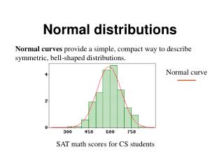
Normal. COPD. 0. 250. Frequency (Hz). Ratio Of Low Frequency Energy To High Frequency Energy Of Lung Sounds In Patients With COPD R. Murphy and A. Vyshedskiy, Brigham and Women’s / Faulkner Hospitals, Boston MA. 80 Hz. Purpose. Normal. Results.

E N D
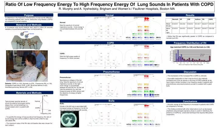
Normal COPD 0 250 Frequency (Hz) Ratio Of Low Frequency Energy To High Frequency Energy Of Lung Sounds In Patients With COPD R. Murphy and A. Vyshedskiy, Brigham and Women’s / Faulkner Hospitals, Boston MA 80 Hz Purpose Normal Results To determine if the frequency characteristics of lung sounds differed in non-wheezing patients with chronic obstructive lung disease (COPD) as compared to normal subjects. Normal: Note the spectrum of normal inspiratory sounds – most energy is concentrated between 80 and 200 Hz. Materials and Methods A 16-channel lung sound analyzer was used to collect 20 seconds samples of sound during deeper than normal breathing. • Notice that R4 was significantly greater in COPD as compared to normals (p<0.05). COPD Frequency Distribution of R4 Age matched COPD (n=128) and Normals (n=128) COPD: Note the high power peaks at frequency 0 to 80Hz (arrows) Pneumothorax Discussion • The mechanism of the increased R4 in COPD is unknown. • A possible explanation is that it may be due to the relatively increased size of the air spaces in the lung of COPD patients as we have noted a similar increase in low frequency peaks in patients with pneumothorax and pneumonectomy as well in a patient with a giant bulla. Pneumothorax: Spontaneous collapse of the left lung. Note the spectrum of normal inspiratory sounds on the right – most energy is concentrated between 80 and 200 Hz. On the left note the presence of a very low frequency peak at 20Hz (arrow) and the absence of energy at higher frequencies. Patients: COPD (n=103), Normals (n=379), Pneumonia (PN, n=118), Congestive heart failure (CHF, n=92), Bronchial asthma (n=62), Interstitial pulmonary fibrosis (IPF, n=39) Materials and Methods Bulla Conclusions Typical power spectral density of sound recorded at lung bases during deeper than normal breathing in a normal subject and in a patient with COPD. Bulla: A bulla in the left lung is associated with increased energy at frequency 0 to 80 Hz and decreased vesicular breath sounds. • Acoustic energy at low frequency is increased in patients with COPD as compared to normals. • This finding combined with other observations such as decreased amplitude and prolonged expiratory phase is useful in identifying the presence of COPD by a bed-side technique that requires little patient cooperation. • To quantify the energy of lung sounds at low frequency, the ratio of sound energy from 20 Hz to 80Hz to that from 80 to 800 Hz was calculated (R4). • The maximum value of the R4 ratio at 8 basilar sites was chosen for each subject.