Download
1 / 34
340 likes | 482 Views
Lecture 3-Amino Acid & Protein. Ahmad Razali Ishak Department of Environmental Health Faculty of Health Sciences UiTM Puncak Alam. Function of Protein. Catalysts- enzymes for metabolic pathway Storage and transport- myoglobin and hemoglobin Structural- actin, myosin

E N D
Lecture 3-Amino Acid & Protein Ahmad Razali Ishak Department of Environmental Health Faculty of Health Sciences UiTM Puncak Alam
Function of Protein • Catalysts- enzymes for metabolic pathway • Storage and transport- myoglobin and hemoglobin • Structural- actin, myosin • Decoding information- translation and gene expression • Hormones and hormone receptors • Specialized functions-antibodies
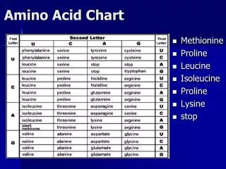
Amino Acids- Primary structure of protein • Amino acid:a compound that contains both an amino group and a carboxyl group • -Amino acid has an amino group attached to the carbon adjacent to the carboxyl group • -carbon also bound to side chain group, R • R gives identity to amino acid
Cont.. • The - carbon is chiral/asymmetric (4 different groups are attached to the carbon: exception is glycine )- mirror image, non super imposable • AA exist as stereoisomer (same molecular formula, but differ in arrangement of groups) – designated D (right) or L (Left) • Vast majority of -amino acids have the L-configuration at the -carbon (Proline is usually D)
-amino group orientation determines L or D • NH3+ on left = L • NH3+ on right = D • Carboxylate group at top- point away side chain at the bottom
AA structure and properties • AA are grouped based upon the properties and structures of side chains • Aliphatic (R groups consist of carbons and hydrogens)-Glycine, Alanine, Valine, Leucine, Isoleucine, Proline
Aromatic (R group have phenyl ring)- Phenylalanine, tyrosine
Cont.. • Basic R group- Histidine, lysine, arginine • Acidic R group - Glutamate, Aspartate • Side chain with alcohols – Serine, threonine
Ionization of Simple Amino Acids • Amino acids are more complicated than simple weak acids since amino acids have at least 2 ionizing groups. • Glycine (abbrevation is Gly), for example, has both a carboxylic acid and an amino group that can ionize:
If we dissolve the free base of Gly in pure water (ie neutral pH), it will ionize. The equilibrium is far to the right so most of the Gly is in the charged form called the Zwitterion and Gly is still neutral because the + charge is neutralized by the - charge. Gly is always in the Zwitterion form at neutral pH.
Ionization of Amino Acids • Remember, amino acids without charged groups on side chain exist in neutral solution as zwitterions with no net charge
If one aa being titrated from acidic condition, you will get this titration curve • E.g. Titration curve for glycine
At given pH, amino acid have different net charge • The isoelectric point (pI) is the pH at which the amino acid has no net charge = zwitterion • If pH > pI, amino acid would be –ve charged • If pH < pI, amino acid would be positively charged
Isoelectric pH • Isoelectric pH, pI: the pH at which the majority of molecules of a compound in solution have no net charge • the pI for glycine, for example, falls midway between the pKa values for the carboxyl and amino groups • Isoelectric pH values for the 20 protein-derived amino acids are given in Table 3.2
Protein Structure Four Levels of Protein Structure: 1. Primary Structure- Polypeptide backbone- Linear sequence of amino acid 2. Secondary Structure- regular patterns formed by primary structure folding-Local Hydrogen bonds along the backbone 3. Tertiary structure- Completely folded polypeptide with one or more domains. Long distance bonding involving the AA side chains 4. Quaternary structure- Association of multiple polypeptides. Protein interactions leading to formation of dimers, tetramers, etc.
Protein Covalent Structure (Protein Primary Structure) I. Peptide Bonds, Peptides and Proteins • Proteins are sometimes called Polypeptides, since they contain many Peptide Bonds
The peptide bond is an amide bond Water is lost in forming an amide bond.
Peptides = Mini-Proteins A pentapeptide -- GlyAlaSerPheGln 1st amino acid is always written on the left and called the Amino terminal, since it is always the only amino acid of the peptide with a free alpha-amino group. Last amino acid is always written on the right and called the Carboxyl terminus, since it is always the only amino acid of the peptide with a free alpha-carboxylic acid group.
SECONDARY STRUCTURE OF PROTEINS • In 1950's, Linus Pauling named the first structures he found by X-ray diffraction, the Alpha Helix and the second structure he found was called Beta Sheet • The 2 COMMON Types of Protein Secondary Structure: • Alpha-helix • Beta-sheet
-Helix • Coil of the helix is clockwise or right-handed • There are 3.6 amino acids per turn • Repeat distance is 5.4Å • Each peptide bond is s-trans and planar • C=O of each peptide bond is hydrogen bonded to the N-H of the fourth amino acid away • C=O----H-N hydrogen bonds are parallel to helical axis • All R groups point outward from helix
-Pleated Sheet • Polypeptide chains lie adjacent to one another; may be parallel or antiparallel • R groups alternate, first above and then below plane • Each peptide bond is s-trans and planar • C=O and N-H groups of each peptide bond are perpendicular to axis of the sheet • C=O---H-N hydrogen bonds are between adjacent sheets and perpendicular to the direction of the sheet
-Pleated Sheet (Cont’d) -bulge- a common nonrepetive irregular 2˚ motif in anti-parallel structure
Myoglobin • A single polypeptide chain of 153 amino acids • A single heme group in a hydrophobic pocket • 8 regions of -helix; no regions of -sheet
Quaternary Structure • Quaternary (4°) structure: the association of polypepetide monomers into multisubunit proteins • dimers • trimers • tetramers • Noncovalent interactions • electrostatics, hydrogen bonds, hydrophobic
Hemoglobin (Hb) • A tetramer of two -chains (141 amino acids each) and two -chains (153 amino acids each); a2b2
Homework • Describe the difference between alpha-helix and beta-sheet protein structures. • Describe the metabolic disorder of protein- phenylketonuria • Please upload the answer in the i-discuss before next seminar