Download
1 / 10
160 likes | 788 Views
LEISHMANIASIS. Aswad H. Al.Obeidy FICMS, FICMS GE&Hep Kirkuk General Hospital. LEISHMANIASIS. Caused by unicellular flagellate intracellular protozoa belonging to the genus Leishmania It comprises several diverse clinical syndromes

E N D
LEISHMANIASIS Aswad H. Al.Obeidy FICMS, FICMS GE&Hep Kirkuk General Hospital
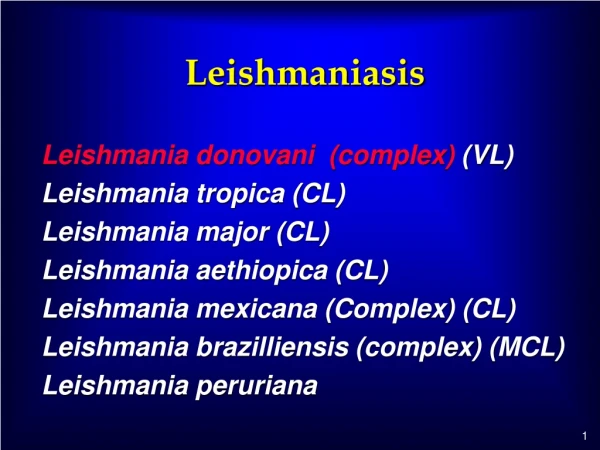
LEISHMANIASIS • Caused by unicellular flagellate intracellular protozoa belonging to the genus Leishmania • It comprises several diverse clinical syndromes • visceral leishmaniasis(VL, kala-azar), the reticuloendothelial system • cutaneous leishmaniasis (CL) , the dermis • mucosal leishmaniasis (ML),the naso-oropharyngeal mucosa • Most clinical syndromes are caused by zoonotic transmission • From animals (chiefly canine and rodent reservoirs) to humans through phlebotomine sandfly vectors • 30 species of phlebotomine sandflies [Phlebotomus (Old World) and Lutzomyia (New World) • Humans are the only known reservoir (anthroponotic) in major VL foci in India and Sudan • There are 21 leishmanial species responsible for these disorders (Leishmania and Viannia) • The disease occurs in 88 countries around the world • Annual incidence of 2 million new cases (500 000 for VL, and 1.5 million for CL)
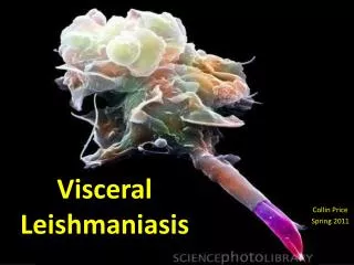
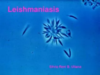
Life cycle • Leishmania parasites, target and persist in tissue macrophages • Flagellar promastigotes (10-20 μm) int. by the feeding female sandfly • Sandfly saliva helps Leishmania evade immunity, sandfly salivary components with immunomodulating effects have been shown to promote experimental infection • Promastigotes bind to receptors on macrophages, are phagocytized, and transform within phagolysosomes into nonflagellated amastigotes, which replicate and infect additional macrophages • Amastigotes(2-4 μm) ingested by sandflies transform back into infective promastigotes • Other modes of transmission include congenital and parenteral (e.g., by blood transfusion or needle sharing). • Flagellar promastigote which multiplies by binary fission in the gut of the vector and migrates to the proboscis to infect a new host.
VISCERAL LEISHMANIASIS (KALA-AZAR) • Caused by the protozoon Leishmania donovani complex • Transmitted by the phlebotomine sandfly • Transmission has also been reported to follow blood transfusion in northern Europe • Can present unexpectedly in immunosuppressed patients-for example, after renal transplantation and in AIDS • A broad spectrum of severity and manifestations, with a chronic, subacute, or acute onset • An incubation period of weeks, months, or sometimes years • The term kala-azar typically is reserved for advanced, life-threatening disease • kala-azar means black [kala] fever [azar] in Hindi, darkening of the skin is uncommon • India, Sudan, Bangladesh and Brazil account for 90% of cases of VL
Clinical features • On the Indian subcontinent both adults and children are equally affected • On other continents it is predominantly a disease of small children and infants, except in HIV co-infection (adult disease). • Malnutrition being both a risk factor for and a sequela of visceral • The first sign of infection is high fever, usually accompanied by rigor and chills. • Fever intensity decreases over time and patients may become afebrile for intervening periods ranging from weeks to months. • This is followed by a relapse of fever, often of lesser intensity. • Hepatosplenomegaly (with splenomegaly usually predominant and the spleen sometimes massive • Anemia; leukopenia (neutropenia, marked eosinopenia, and relative lymphocytosis and monocytosis); thrombocytopenia. Pancytopenia with its consequent clinical manifestations is a common feature • Hypergammaglobulinemia (chiefly IgG, from polyclonal B cell activation); and hypoalbuminemia. • Blackish discoloration of the skin is a feature of advanced illness
Complications • Profound immunosuppression and secondary infections are very common • Tuberculosis • Pneumonia • Severe amoebic or bacillary dysentery • Gastroenteritis • Herpes zoster and chickenpox • Skin infections, boils, cellulitis and scabies are common • Without adequate treatment most patients with clinical VL are likely to die.
Differential diagnosis • Typhoid fever • Subacute bacterial endocarditis • Miliary tuberculosis • Brucellosis • Histoplasmosis • Tropical splenomegaly syndrome • Schistosomiasis • Myeloproliferative diseases (e.g., leukemia and lymphoma).
Investigations • Pancytopenia is the most dominant feature, granulocytopenia and monocytosis • Polyclonal hypergammaglobulinaemia, chiefly IgG followed by IgM, and hypoalbuminaemia • Patients are anergic to the leishmanin antigen skin test (LST) • Demonstration of amastigotes (Leishman-Donovan bodies) in splenic smears is the most efficient means of diagnosis, with 98% sensitivity • Safer methods like bone marrow or lymph node smears are not as sensitive • PCR for DNA detection from the peripheral blood is an efficient non-invasive method for diagnosis • Serodiagnosis, by ELISA or immunofluorescence antibody test, is employed in developed countries • Direct agglutination test of stained promastigotes and an equally efficient rapid immunochromatographic k39 strip test
Management • Pentavalent antimonials, first drugs used for the treatment of leishmaniasis and remain the mainstay of treatment, is available as sodium stibogluconate (100 mg/ml).The daily dose is 20 mg/kg body weight, given either intravenously or intramuscularly for 28-30 days. Side-effects are common and include arthralgias, myalgias, raised hepatic transaminases, pancreatitis, especially in patients co-infected with HIV, and ECG changes • Amphotericin B deoxycholate given once daily or on alternate days at a dose of 0.75-1.00 mg/kg for 15-20 doses, It has a cure rate of nearly 100%. • Miltefosine,this oral drug,a daily dose of 50 mg (patient's body weight < 25 kg) to 100 mg (≥ 25 kg), or 2.5 mg/kg body weight for children, for 28 days cures over 90% of patients. • Paromomycin,an aminoglycoside, given intramuscularly at 15 mg/kg body weight daily for 3 weeks. • Pentamidine isetionate, declining efficacy and serious side-effects such as type 1 diabetes mellitus, hypoglycaemia and hypotension have led to it being abandoned.
Post-kala-azar dermal leishmaniasis (PKDL) • After treatment and recovery, some patients develop dermatological manifestations in a small minority of patients 6 months to ≥ 3 years after the initial infection • as macules, papules, nodules (most frequently) and plaques which have a predilection for the face • Hypopigmented macules can occur over all parts of the body • There are no systemic symptoms and no spontaneous healing • The diagnosis is clinical, supported by demonstration of scanty parasites in lesions by slit skin smear and culture • Immunofluorescence and immunohistochemistry are other met. • Serological tests (direct agglutination test or k39 strip tests) are positive. • Treatment of PKDL is difficult. Sb for 120 days or several courses of amphotericin B infusions • PKDL patients are a human reservoir, and focal outbreaks have been linked to patients with PKDL