Download
1 / 42
420 likes | 605 Views
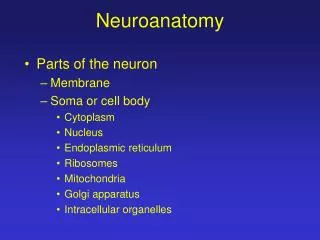
Lecture 4-Neuroanatomy Walter Schneider When we talk about the brain we need to be able to identify and communicate clearly on what part of the brain we are discussing (slides contributed by Julie Fiez (U. Pittsburgh) & Rainer Goebel U. Maastricht, Henk Jansma (Brain Innovation).

E N D
Lecture 4-Neuroanatomy Walter Schneider When we talk about the brain we need to be able to identify and communicate clearly on what part of the brain we are discussing (slides contributed by Julie Fiez (U. Pittsburgh) & Rainer Goebel U. Maastricht, Henk Jansma (Brain Innovation)
How was each slice made? What major distinctions can you see? What do you think that they represent?
What happens if we “zoom” in on the grey matter? How can we visualize the structure more clearly? What features can we observe?
Is every piece of the cortex the same? How might different pieces of the brain be different?
What did Joseph Gall (1758-1828) have right and wrong? How should this make today’s cognitive neuroscientists careful about what they say.
Brain Stretching and RotatingSpatial normalization – why? Single subject level: Integrating the results of multiple recording sessions of the same subject -> increased statistical power Multiple subject level: Comparing results across different subjects, summarizing results at the group level Attempt to generalize statistical analysis to the investigated group (fixed effects analysis) or to the population level (random effects). Statistics „runs“ over voxels of different subjects. Running statistics over subjects is statistically effective only if spatial correspondence between brain areas has been established.
Spatial mapping: approaches Spatial transformation of brains in Talairach spacePro:Widespread acceptanceCons:Rather crude alignment; requires spatial smoothing, which introduces, however, severe problems like blurring of functionally distinct areas and the suppression of significant small areas Region of Interest (ROI)-based alignment: Identification of functional areas by premapping experiments (e.g., FFA, PPA, V1,V2)Pro:Optimal statistical analysis possibleCons:Not easily applicapple in complex cognitive tasks, focuses on „known“ areas, not automatic
Talairach reference system • Talairach transformation uses 8 anatomical landmarks of a human brain for spatial normalization • Anterior Commissure AC • Posterior Commissure PC • Front AP • Back PP • Top SP • Bottom IP • Left • Right
Defining 8 Anatomical Landmarks • anterior commissure AC • posterior commissure PC • anterior point AP • posterior point PP • superior point SP • inferior point IP • right point RP • left point LP
Identifying Anterior and Posterior Commissure • This is a reasonably easy to find location viable on anatomical scans providing a 0,0,0 mark for translation within the head Z Z X Y Y X
Anterior Posterior CommissureAxial View DV 2.6 DV 0.0
Flowchart for Talairach transformation Talairach step 1 Talairach step 2 ACPC- rotation Borders of cerebrum
Cortex Landmarking Analogy identifying Pittsburgh as North America, Pennsylvania, Allegany County, Latitude: 40° 25.53' N Longitude: 79° 58.062' W, on the intersection of the Ohio, Monongahela and Allegany rivers, Intersection of I279 & I376, Old steel production center • What kind of labels/landmarking are useful? • How difficult are they to achieve reliably? • How would the be used by different scientific communities
Cortex Landmarking • Lobes – general areas • Talairach – X,Y, Z location • Gyri + Sulci – surface features • Brodmann – anatomical numbering of areas • Area numbering – based on multiple anatomical and or functional metrics (e.g., in vision V1, V2, V3, V3A, V4, TEO, TE MT)
Notes on Learning Anatomy • BrainTutor • Human Brain Coloring Book • Digital Atlas • Physical Models • Talairach Atlas • Other Atlases • NMI brain
Cortical Areas Brain Tutor – Goebel Damasio
Brodmann Areas with BrainTutor Brain Tutor – Goebel Damasio
BrainTutor Learning Anatomy V 2.0 V 1.0
Homework Digital Coloring of Brain Example overlay on postcentral gyrus Example overlay on postcentral gyrus
Digital Anatomist http://www9.biostr.washington.edu/da.html
Digital Anatomist Cross Check http://www9.biostr.washington.edu/cgi-bin/DA/imageform
Talairach Daemon Cross Check http://ric.uthscsa.edu/td_applet/
Example of Brain Atlases Talairach MNI Montreal Neuro. Inst. Structural Image http://www.bic.mni.mcgill.ca/cgi/icbm_view/
Summary • It is important to accurately communicate and encode locations in the brain. • There are multiple coding schemes (Talairach, Brodmann, Gyri + Sulci, ROI based (e.g., V1, FFA). • You need to be able to understand all these methods. • There are good tools available to idenify structures (particularly BrainTutor and Digital Anatomist). • Note labeling takes some time to develop skill and there are areas of ambiguity.