Download
1 / 96
1.27k likes | 2.24k Views
Cardiac Arrhythmias. Dr. Ahmad Hersi. Myocardium Muscle Action Potential. Normal Cardiac Cycle. What does it tell us?. the electrical conduction through the heart areas of ischemia or myocardial damage LV Hypertrophy electrolyte disturbances / drug toxicity.

E N D
Cardiac Arrhythmias Dr. Ahmad Hersi
What does it tell us? • the electrical conduction through the heart • areas of ischemia or myocardial damage • LV Hypertrophy • electrolyte disturbances / drug toxicity
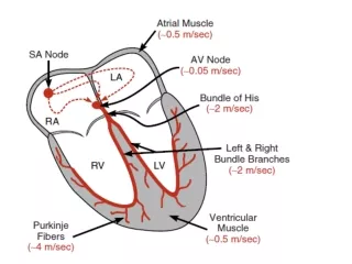
The Electrical System of the Heart Inter- nodal Tracts SA Node Left Bundle Branch AV Node Anterior Superior Fascicle Bundle of HIS Posterior Inferior Fascicle Septal Depolarization Fibers Purkinjie Fibers Right Bundle Branch
Conduction System of the Heart: A Conceptual Model for Illustration Inter-nodal Tract Left Bundle Branch AV Node Septal Depolarization Fibers Bundleof HIS SA Node James Fibers Anterior Superior Fascicle Posterior Inferior Fascicle Right Bundle Branch Bundle of Kent
Inter- nodal Tracts SA Node SA Node – “pacemaker” of the heart (60-100bpm) AV Node – junction of the atria and ventricles (40-60bpm) Bundles – Bundle of His connects the AV node to the bundle branches (20-40bpm) AV Node Bundle of HIS
What Is In Each Beat? (the cardiac cycle in waves, complexes, and intervals) • P Wave – atrial contraction or depolarization, (usually upright) • QRS Complex – time for ventricular contraction or depolarization (usually upright) (0.04 - 0.12sec) (delays in the bundle branches will widen the QRS) • T Wave – ventricular repolarization “recharging” (usually upright) • PR Interval – time between atrial depolarization to ventricular depolarization (beginning of P wave to beginning of QRS)(0.12 - 0.20sec) (prolonged PR = delays in the AV node conduction) • QT Interval – represents one complete ventricular depolarization and repolarization (beginning of QRS to the end of the T wave) (0.32 – 0.44sec) (disturbances are usually due to electrolyte disturbances or drug effects)
ECG Paper and related Heart Rate & Voltage Computations Memorize These 2
Reading a Rhythm StripWhat Do I Look For? • Regularity - What is the R – R Interval? • Rate - Is the rate normal (60-100), slow, or fast? ***Six-second strip method - (30 big boxes) & multiply times ten • P Wave– Is there a P wave before every QRS? Is it upright? • QRS Complex– Is there a normal QRS complex following each P wave? Wide or normal? • T wave– How does your T wave look? Upright? • Measure your intervals– PR Interval, QRS, QT
Pacemakers of the Heart • SA Node - Dominant pacemaker with an intrinsic rate of 60 - 100 beats/minute. • AV Node - Back-up pacemaker with an intrinsic rate of 40 - 60 beats/minute. • Ventricular cells - Back-up pacemaker with an intrinsic rate of 20 - 45 bpm. Tehran Arrhythmia Center
Rhythm Analysis • Step 1: Calculate rate. • Step 2: Determine regularity. • Step 3: Assess the P waves. • Step 4: Determine PR interval. • Step 5: Determine QRS duration. Tehran Arrhythmia Center
Step 1: Calculate Rate 3 sec 3 sec Option 1 • Count the # of R waves in a 6 second rhythm strip, then multiply by 10. Interpretation? 9 x 10 = 90 bpm Tehran Arrhythmia Center
Step 1: Calculate Rate • Option 2 • Find a R wave that lands on a bold line. • Count the # of large boxes to the next R wave. If the second R wave is 1 large box away the rate is 300, 2 boxes - 150, 3 boxes - 100, 4 boxes - 75, etc. (cont) R wave Tehran Arrhythmia Center
Step 1: Calculate Rate 300 150 100 75 60 50 • Option 2 (cont) • Memorize the sequence: 300 - 150 - 100 - 75 - 60 - 50 Interpretation? • Approx. 1 box less than 100 = 95 bpm Tehran Arrhythmia Center
Step 2: Determine regularity R R • Look at the R-R distances (using a caliper or markings on a pen or paper). • Regular (are they equidistant apart)? Occasionally irregular? Regularly irregular? Irregularly irregular? Interpretation? Regular Tehran Arrhythmia Center
Step 3: Assess the P waves • Are there P waves? • Do the P waves all look alike? • Do the P waves occur at a regular rate? • Is there one P wave before each QRS? Interpretation? Normal P waves with 1 P wave for every QRS Tehran Arrhythmia Center
Step 4: Determine PR interval • Normal: 0.12 - 0.20 seconds. (3 - 5 boxes) Interpretation? 0.12 seconds Tehran Arrhythmia Center
Step 5: QRS duration • Normal: 0.04 - 0.12 seconds. (1 - 3 boxes) Interpretation? 0.08 seconds Tehran Arrhythmia Center
Rhythm Summary • Rate 90-95 bpm • Regularity Regular • P waves Normal • PR interval 0.12 s • QRS duration 0.08 s Interpretation? Normal Sinus Rhythm Tehran Arrhythmia Center
Normal Sinus Rhythm • Normal and constant P wave contours • Normal P wave axis • Rate between 60 and 100 bpm Tehran Arrhythmia Center
Anatomical Aspects of Normal Sinus Node • Located at the superior anterolateral portion of right atrium near its border with the superior vena cava • It is an epicardial structure near sulcus terminalis • From endocardial approach the closest approach is near the superior end of crista terminalis Tehran Arrhythmia Center
Sinus Node Function • The dominant cardiac pacemaker • Highly responsive to autonomic influences • Decreasing rate with vagal stimulation • Increasing rate with sympathetic activity • Normal sinus rate under basal conditions is 60-100 bpm. Tehran Arrhythmia Center
Sinus Tachycardia • Rate? 130 bpm • Regularity? Regular • P waves? Normal • PR interval? 0.16 s • QRS duration? 0.08 s Interpretation? Sinus Tachycardia Tehran Arrhythmia Center
Sinus Tachycardia • Sinus rhythm exceeding 100 bpm in adults • Usually between 100 and 180 bpm but may be higher with extreme exertion • Maximum heart arte decreases wit age from near 200 bpm to less than 140 bpm • Gradual onset and termination Tehran Arrhythmia Center
Sinus Tachycardia Tehran Arrhythmia Center
Sinus TachycardiaCauses • Common in infancy and childhood • Normal response to a variety of physiological and pathological stresses • Exertion, anxiety • Hypovolemia, anemia • Fever • Congestive heart failure • Myocardial ischemia • Thyrotoxicosis • Drugs • Inflammation Tehran Arrhythmia Center
Sinus Bradycardia • Rate? 30 bpm • Regularity? Regular • P waves? normal • PR interval? 0.12 s • QRS duration? 0.10 s Interpretation? Sinus Bradycardia Tehran Arrhythmia Center
Sinus Bradycardia • Sinus rhythm at a rate less than 60 bpm • Can result from excessive vagal or decreased sympathetic tone as well as anatomic changes in sinus node • Frequently occurs in healthy young adults, particularly well-trained athletes • Sinus arrhythmia often coexists Tehran Arrhythmia Center
Sinus Bradycardia Tehran Arrhythmia Center
Sinus BradycardiaJunctional Escape Beats Tehran Arrhythmia Center
Sinus Bradycardia Causes • Hypothyroidism • Drugs • During vomiting or vasovagal syncope • Increased intracranial pressure • Hypoxia, hypothermia • Depression • Jaundice Tehran Arrhythmia Center
Sinus Arrhythmia • Rate? 50-75 bpm • Regularity? Phasic variations • P waves? normal • PR interval? 0.12 s • QRS duration? 0.10 s Interpretation? Sinus Arrhythmia Tehran Arrhythmia Center
Sinus Pause Tehran Arrhythmia Center
Sick Sinus Syndrome A combination of symptoms (dizziness, fatigue, confusion, syncope and congestive heart failure) caused by sinus node dysfunction Atrial tachyarrhythmias may accompany sinus node dysfunction <bradycardia-tachycardia syndrome> Tehran Arrhythmia Center
AV BlockTypes • First degree AV block • Second degree AV block • Mobitz type I (Wenckebach) • Mobitz type II • Third degree AV block (Complete heart block) • High degree (advanced) AV block Tehran Arrhythmia Center
First Degree AV Block • Rate? 60 bpm • Regularity? Regular • P waves? Normal • PR interval? 0.36 s • QRS duration? 0.08 s Interpretation? 1st Degree AV Block Tehran Arrhythmia Center
PR Interval PR interval < 0.12 s 0.12-0.20 s > 0.20 s High catecholamine states Wolff-Parkinson-White Normal AV nodal blocks Wolff-Parkinson-White 1st Degree AV Block Tehran Arrhythmia Center
First Degree AV Block • Conduction time is prolonged but all impulses are conducted. • PR interval exceeds 0.2 sec in adults • Site of conduction delay may be in the AV node (most commonly), in the His-Purkinje system or both. Tehran Arrhythmia Center
First Degree AV Block Tehran Arrhythmia Center
Wenckebach AV Block • Rate? 50 bpm • Regularity? Regularly irregular • P waves? Nl, but 4th no QRS • PR interval? Lengthens • QRS duration? 0.08 s Interpretation? 2nd Degree AV Block, Type I Tehran Arrhythmia Center
Mobitz Type I Second Degree AV Block • Also called Wenckebach block • Typical type characterized by progressive PR prolongation culminating in a non-conducted P wave • Narrow QRS in most cases Tehran Arrhythmia Center
WB Tehran Arrhythmia Center
Wenckebach Block • Atypical pattern in over half the cases • The site of block is almost always in the AV node. • Generally benign and does not advance to more advanced AV block • Can occur in normal children and well-trained athletes Tehran Arrhythmia Center
Mobitz Type II AV Block • Rate? 40 bpm • Regularity? Regular • P waves? Nl, 5th P no QRS • PR interval? 0.18 s • QRS duration? 0.11 s Interpretation? 2nd Degree AV Block, Type II Tehran Arrhythmia Center
Mobitz Type II Second Degree AV Block • PR interval remains constant prior to the blocked P wave • Commonly associated with bundle branch blocks Tehran Arrhythmia Center