Download
1 / 19
230 likes | 905 Views
hemoglobin. Dr. Dalia Kamal Eldien. Structure of hemoglobin.

E N D
hemoglobin Dr. Dalia KamalEldien
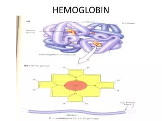
Structure of hemoglobin • Hemoglobin abbreviated as (Hb), the main component of the red blood cell, is a conjugated protein that serves as the vehicle for the transportation of oxygen and carbon dioxide. When fully saturated, each gram of hemoglobin holds 1.34ml of oxygen. The red cell mass of the adult contains approximately 600g of hemoglobin, capable of carrying 800ml of oxygen.
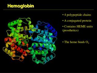
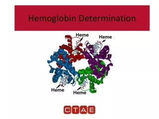
A molecule of hemoglobin consists of two pairs of polypeptide chains (globin) and four heme groups, each containing one atom of ferrous iron. Each heme group is precisely located in a pocket or fold of one of polypeptide chains. Located near the surface of the molecule, the heme reversible combines with one molecule of oxygen or carbon dioxide.
At least three distinct hemoglobin types are found postnatally in normal individuals, and the structure of each has been determined • Hb A • HbA2 • Hb F
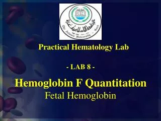
Hb A is the major (96-98%) normal adult hemoglobin. The polypeptide chains of the globin part of the molecules are of two types: two identical α-chains, each with 141 amino acids; and two identical β-chains, with 146 amino acids each. Hb F is the major hemoglobin of the fetus and the new born infant. The two α-chains are identical to those of Hb A; and two γ-chains, with 146 amino acids residues, differ from β-chains. Only traces of Hb F (<1.0%) are found in adults. Hb A2 account for 1.5% to 3.5% of normal adult hemoglobin. Its two α-chains are the same as in Hb A and Hb F; its two δ-chains differ from β-chains in only 8 of their 146 amino acids.
Hemoglobin (Hb) is synthesized in a complex series of steps. The heme part is synthesized in a series of steps in the mitochondria and the cytosol of immature red blood cells, while the goblin protein parts are synthesized by ribosome's in the cytosol. Production of Hb continues in the cell throughout its early development from the proerythroblast to the reticulocyte in the bone marrow. At this point, the nucleus is lost in mammalian red blood cells, but not in birds and many other species. Even after the loss of the nucleus in mammals, residual ribosomal RNA allows further synthesis of Hb until the reticulocyte loses its RNA soon after entering the vasculature
Heme synthesis: Occurs in most cells of the body, except the mature erythrocytes, but most abundantly in the erythroid precursors. Succinyl-coenzyme condenses with glycine to form the unstable intermediate α-amino β-ketoadipic acid, which is readily decarboxylated to δ-aminolevulinic acid (ALA). This condensation requires pyridoxal phosphate (vitamin B6) and occurs in mitochondria. 2 molecules of ALA condense to form the monopyrrole, porphobilinogen. 4 molecules of porphobilinogen react to form uroporphyrinogen III or I. Type III isomer is converted to protoporphyrin IX. Iron is inserted into protoporphyrin by the mitochondrial enzyme ferrochetalase to form the finished heme moiety.
Globin synthesis: • Occurs in the cytoplasm of the normoblast and reticulocyte. The polypeptide chains are manufactured on the ribosomes. Progressive growth of the polypeptide chain begins at the amino end. This process of protein synthesis occurs on ribosomes clustered into polyribosomes, which are held together by the mRNA. The polypeptide chains released from the ribosomes are folded into their three-dimensional configurations spontaneously. The complete globin structure consists of four polypeptide chains formed by two dissimilar pairs.
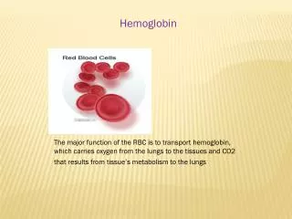
Function of hemoglobin • The functions of hemoglobin include: • Transport of O2 from the lungs to the tissues and of CO2in the reverse direction. • Assisting in acid-base regulation by eliminating CO2 in the lungs and by the buffering action of hemoglobin.
Hemoglobin is measured to detect anemia and its severity and to monitor an anemic patient’s response to treatment. The test is also performed to check the hemoglobin level of a blood donor prior to donating blood. Capillary blood or EDTA anticoagulated venous blood can be used. Hemoglobin values are expressed in grams per liter (g/l) or grams per deciliter (g/dl). Grams/liter is the recommended way of expressing the mass concentration of hemoglobin.
The hemoglobin content a solution may be estimated by several methods: • measurement of color • measure the power of combining with oxygen or carbonmonoxide • Measure the iron content. • Hemoglobin is measured photometrically or estimated using a visual comparative technique. In photometric techniques the absorbance of hemoglobin in a blood sample is measured electronically using a filter colorimeter or a direct read-out hemoglobin meter. When it is not possible to measure hemoglobinaccurately using a photometric technique; a visual comparative technique can help to detect anemia and assess its severity.
Estimation methods are: Cyanmethemoglobin (hemiglobincyanide- hicn) method. Hemocue non-dilution photometric technique. Oxyhemoglobin method. Alkaline hematin method. Acid hematin method (sahli-hellige) . Hemoglobin color scale. Copper sulphate densitometery.
Interpretation of hemoglobin test results: Normal hemoglobin levels vary according to age and gender, and the altitude at which a person lives. Normal hemoglobin reference range: Children at birth 135-195 g/l children 2 y – 5 y 110-140 g/l Children 6 y – 12 y 115-155 g/l Adult men 130-180 g/l Adult women 120-150 g/l Pregnant women 110-138 g/l