Download

1 / 120
1.2k likes | 1.2k Views
Learn about the epidemiology, assessment, and management of burns, including immediate and definitive treatment, complications, and special considerations. Explore the different types of burns, their effects, and the phases of burn care.

E N D
BURNS Dr. Neil D’Souza
Overview • Epidemiology • Definition • Assessment • Management • Immediate • Definite • Complications • Special considerations
Epidemiology • More then 6 million people are burned in India every year • Most are minor burns and treated in outpatient • About 5% require hospitalization for appropriate treatment
Death in burns is a typical bimodal distribution • Immediately after injury • Weeks later as a result of multi-organ failure • 2/3rd of burns occur at home involving children less then 15yrs or elderly more than 60yrs
Definition of a Burn “Tissue injury caused by thermal, radiation, chemical, or electrical contact resulting in protein denaturation, burn wound edema, and loss of intravascular fluid volume due to increased vascular permeability.”
Approach to a Patient with Burns • Cause of the burns • Assessment of burn • Depth • Extent • Immediate casualty treatment
Definitive treatment • Fluid resuscitation • Nutrition • Treatment of wound • Infection • Surgical intervention • Prevention of complications and their treatment
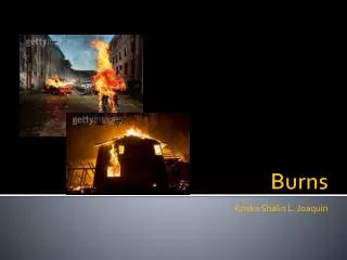
Types of Burns • Thermal burns • Scald • Flame • Flash • Contact • Electrical burns • Chemical burns • Cold injury • Radiation
Effects of burns influenced by • Intensity • Duration of exposure • Type of tissue
Classification of burns • Depending on thickness of skin involved • Depending on percentage of burns
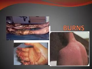
Classification • First degree • Second degree • Third degree • Fourth degree
First-Degree Burns • Does not go below basal layer of the epidermis • Dry and painful • Appears red due to increased blood flow • Heals in a few days
Second-Degree Burns • Extends below basal layer, but not completely through dermis • Superficial • Blister, very painful, contains skin parts (adnexa) which assist in epithelialization • Deep Partial-thickness • Deeper in dermis, fewer adnexa, longer healing time, less painful
Third-Degree Burns • Extends completely through dermis • Adnexa destroyed so can’t heal by epithelialization • Dermal plexus of nerves destroyed-less painful • Burns can be yellow, red, black, brown
Fourth-degree burns • Full-thickness destruction of skin/subcutaneous tissue • Involves underlying fascia, muscle, bone or other structures • Prolonged disability
Mild • Partial thickness < 15% in adult or <10% in children • Full thickness < 2% • Treated as outpatient
Moderate • Partial thickness 15-20% • Full thickness 2-10% • Severe • Partial thickness > 25% • Full thickness > 10% • Involving eyes, ears, feet, hands, perineum • Burns with trauma
Zones of Burn Injury • Zone of Coagulation • Inner Zone • Area of cellular death (necrosis) • Zone of Stasis • Area surrounding zone of coagulation • Cellular injury: decreased blood flow & inflammation • Potentially salvable; susceptible to additional injury • Zone of Hyperemia • Peripheral area of burn • Area of least cellular injury & increased blood flow • Complete recovery of this tissue likely.
Indications for admission • Moderate or severe burns (2nd or 3rd degree) • Airway burns • Burns in extremes of age • Electrical or deep chemical burns • Burns with significant co-morbid conditions • Burns in pts who require special emotional, social intervention
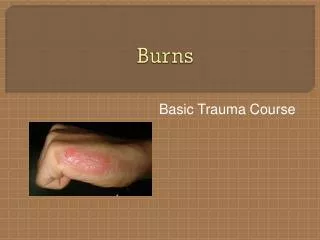
Presentation • H/o Burn • Pain • Anxious state • Blisters • Tachycardia • Tachypnea • In severe cases, shock
Fluid shift • Vessels adjacent to burn injury dilate → ↑ capillary hydrostatic pressure and ↑ capillary permeability • Continuous leak of plasma from intravascular space into interstitial space • Associated imbalances of fluids, electrolytes and acid-base occur • Hemoconcentration • Lasts 24-36 hours
After 36 hrs, fluid leak ceases • Fluid shifts back into circulation • Restores fluid balance and renal perfusion
Inflammatory reaction is localised in small burns • After 10-15% TBSA burns, inflammatory reaction (fluid loss) can lead to shock • Volume of fluid lost is directly proportional to area of the burn
Effects of burns • Shock • Renal failure • Respiratory distress • Infections • Erosive gastritis • Electrolyte imbalance • Immunosuppression
Phases of Burn Care • Emergency care (ABCs) • Resuscitation (hours 0-48) • Definitive care (day 3 until wounds are closed) • Rehabilitation
First Aid • Keep the patient away from the source • Clothing to be removed • Clean the part • Cool the area with tap water • Cover the part
ABCs of Emergency Burn Care (Advanced Burn Life Support) • A = Airway (with cervical spine assessment) • B = Breathing • C = Circulation • D = Disability • E = Exposure and Environmental Control • F = Fluid Resuscitation based on Burn Size and Weight Measurement • Secondary Survey
Definitive Treatment • Fluid resuscitation • Local management
Fluid Resuscitation • 1 or 2 large bore IV lines • Fluid replacement based on: size/depth of burn, age of pt. • Palmar Method • Rule of Nines • Lund-Browder Method • Formula’s for replacement: Parkland formula and Brooke formula
Nutrition • Burns patients need more calories • Early enteral feeding in pts > 20 TBSA burns
Local management • 1st degree burns • Regular dressings is mainstay • Topical ointments like neosporin , povidone iodine will suffice
2nd degree burns • Regular dressings with antibiotic ointments • Silver sulfadiazine • Mafenide acetate • Silver nitrate • Or temporary coverage using biological/artifical synthetic coverings • If 2nd degree burns don’t heal within 2 weeks, skin grafting is indicated
Biological/artifical coverings • Biological • Autograft • Homograft • Heterograft (xenograft) • Isograft • Amniotic membrane • Cultured skin
Artificial skin • Two layered which creates an artificial dermis • Synthetic dressing • Solid silicone and plastic dressing • Can see through to monitor wound status
Collagen dressing • Indications • Deep 2nd degree (non infected) • 3rd degree burns as temporary covering after surgery • Advantages • Adheres to raw surfaces • Peels off as wound heals and epithelisation occurs • Promotes healthy granulation tissue in deep wounds
Artificial skin • Complex of collagen and condroiton sulphuric acid with silicon membrane
Systemic antibiotic therapy • Usually not recomended in the first 48 hrs • After 48 hrs, broad spectrum antibiotics started • Re-evaluation with C&S should be done every 5 days • Resistance and superinfection is common
Deep burns ( deep 2nd degree and 3rd degree) • Early excision • Tangential • Sequential • Followed by • SSG • Full thickness graft