Download
1 / 15
230 likes | 687 Views
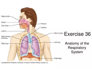
Anatomy: Trachea. Chris van Zyl KHC. Trachea: Landmarks. 2. Begins at lower border of cricoid cartilage / C6 Extends to Carina Right of the midline Sternal angle T4 on inspiration / T6 on expiration Lined by ciliated columnar epithelium 15 cm long / 2cm in diameter

E N D
Anatomy: Trachea Chris van Zyl KHC
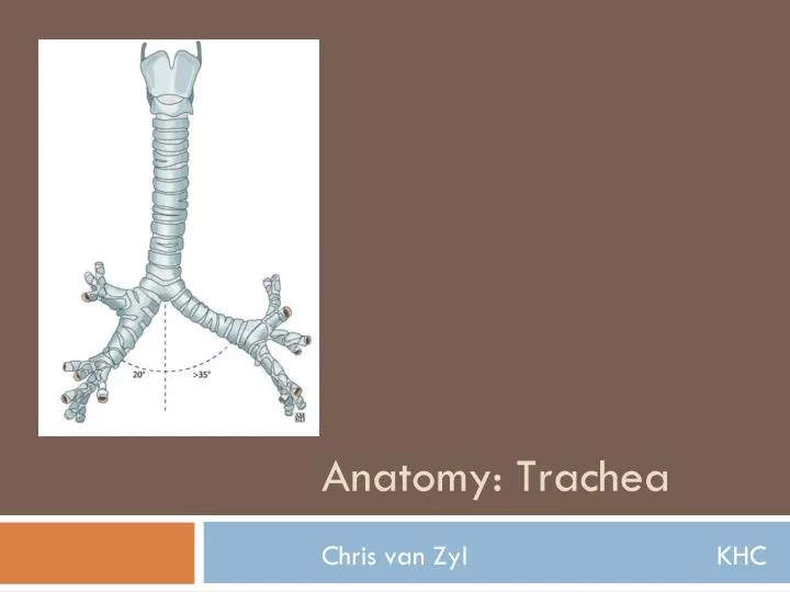
Trachea: Landmarks 2 • Begins at lower border of cricoid cartilage / C6 • Extends to Carina • Right of the midline • Sternal angle • T4 on inspiration / T6 on expiration • Lined by ciliated columnar epithelium • 15 cm long / 2cm in diameter • 15 – 20 incomplete rings of cartilage • Bridged post. by trachealis muscle
Trachea 3
Trachea 4 • Variable shape • Usually round, oval, oval with flattened post. border • Square • Inverted pear • Horseshoe • Very pliable in children • May deviate to the right at almost 90° in normal expiratory film.
Trachea: Carina 5 • Lies at T5 level • T4 on expiration • T6 on inspiration • Normal angle: 65° • 20° to right of midline • 40° to left of midline • Angle slightly larger + symmetrical in children • Angle increases by 10° - 15° in recumbency
Relations: Cervical 6 • Anterior: • Isthmus anterior to 2nd, 3rd, 4th rings • Inferior thyroid veins • Strap muscles: Sternohyoid, Sternothyroid • Posterior: • Oesophagus, recurrent laryngeal nerves • Lateral: • Lobes of thyroid • Common carotid artery
Relations: Thoracic 7 • Anterior: • Brachiocephalic a. • Left common carotid a. • Left brachiocephalic v.
Relations: Thoracic 8 • Posterior: • Oesophagus • Left recurrent laryngeal n.
Relations: Thoracic 9 • Left lateral: • Arch of the aorta • Left common carotid • Left subclavian arteries • Right lateral: • Right vagus nerve • Arch of the azygos vein • Pleura
Blood supply 10 • Upper trachea • Inferior thyroid artery • Lower part • Branches of the bronchial artery • Venous drainage • Inferior thyroid venous plexus
Right Paratracheal Stripe 11 • Interface between trachea and right upper lobe • Max normal thickness 4 mm
Left Paratracheal Stripe 12 • Left upper lobe and trachea • Less common (only on 21 %- 31% of films
Posterior Tracheal Stripe 13 • Interface between trachea and right lung • Normal max diameter 2.5 mm
Tracheoesophageal Stripe 14 • Interface between esophagus and trachea • Normal max diameter 5.5 mm
References 15 • Lines and Stripes: Where Did They Go? • Jerry M. Gibbs, Chitra A. Chandrasekhar, Emma C. Furgasson, • Sandra A. A. Oldham • Applied Radiological Anatomy • Paul Butler, Adam W. M. Mitchell, Harold Ellis • Anatomy for Diagnostic Imaging • Stephanie Ryan, Michelle McNicholas, Stephen Eustace • Third Edition