Download
1 / 57
570 likes | 587 Views
ERYSIPELAS. KAVITA NATHAN Group 318. what is erysipelas ?. It is a streptococcal infection of the superficial lymphatic vessels, usually associated with broken skin on the face. The area affected is erythematous and oedematous.

E N D
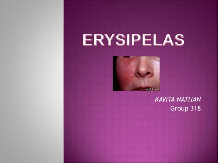
ERYSIPELAS KAVITA NATHAN Group 318
what is erysipelas ? • It is a streptococcal infection of the superficial lymphatic vessels, usually associated with broken skin on the face. • The area affected is erythematous and oedematous. • The patient may be febrile and have a leucocytosis.
pathophysiology Bacteria inoculation into an area of skin, trauma is the initial event in the developing erysipelas
In erysipelas, the infection rapidly invades and spreads through the lymphatic vessels. This can produce overlying skin "streaking" and regional lymph node swelling and tenderness. Immunity does not developto the inciting organism.
RISK FACTORS INCLUDE: • A cut in the skin • Problem with drainage through the veins or lymph system • Skin sores( ulcers)
causes • Streptococcal toxins are thought to contribute to the brisk inflammation that is pathognomonic of this infection. • they clearly coexist with streptococci at sites of inoculation.
Recently, atypical forms reported to be caused by : • * Streptococcus pneumoniae, • *Klebsiella pneumoniae, • * Haemophilus influenzae, • *Yersinia enterocolitica, • *Moraxella species,
Causative agent • *Streptococciare the primary cause of erysipelas. • * Most facial infections are attributed to group Astreptococci, • *lower extremity infections being caused by non–group A streptococci.
Group A beta- hemolytic streptocci • Hemolytic streptococcus • Skin infection • Painful rashes Erythematous rash • Edematous rash • Skin ulcer • Abrasions • Skin ulcer • Insect bite • eczema
symptoms • Blisters • Fever, shaking, and chills • Painful, very red, swollen, and warm skin underneath the sore (lesion) • Skin lesion with a raised border • Sores (erysipelas lesions) on the cheeks and bridge of the nose
Erysipelas begins as a small erythematous patch that progresses to a fiery-red, indurated , tense, and shiny plaque
The lesion classically exhibits raised sharply demarcated advancing margins. • Local signs of inflammation • warmth, • edema, • tenderness are universal.
Lymphatic involvement often is manifested by overlying skin streaking and regional lymphadenopathy
More severe infections may exhibit numerous vesicles and bullae along with petechiae and even frank necrosis.
Signs and test • Erysipelas is diagnosed based on how the skin looks. A biopsy of the skin is usually not needed.
Differential diagnoses • 1) Erythema Annulare Centri-fugum2) Stasis Dermatitis • 3) Cellulitis • 4) Erysipeloid
Erythema annulare centrifugum * Eruptions occur at any age.
Lesions most often appear on the thighs, legs, face, trunk and arms. linked to underlying diseases , viral , bacterial or even tumor.
* acute bacterial infection of traumatized skin. • * caused by Erysipelothrixrhusiopathiae(gram positive rod-shaped bacterium), which cause animal and human infections. • * Direct contact between infected meat and traumatized human skin results in Erysipeloid. • more common among farmers, butchers, cooks, homemakers. • * Lesions most commonly affect the hands.
Antibiotics such as penicillin are used to eliminate the infection. In severe cases, antibiotics may need to be given through an IV (intravenous line). • Those who have repeated episodes of erysipelas may need long-term antibiotics.
Medical care • * Elevation and rest of the affected limb are recommended to reduce local swelling, inflammation, and pain. • * Saline wet dressings should be applied to ulcerated and necrotic lesions and changed every 2-12 hours, depending on the severity of the infection.
*A first-generation cephalosporin or macrolide, such as erythromycin or azithromycin, may be used if the patient has an allergy to penicillin.
Two new drugs: roxithromycin & pristinamycin, have been reported to be extremely effective in the treatment of erysipelas.