Download

1 / 22
240 likes | 607 Views
ANTIBODY DIVERSITY II. Macfarlane Burnet (1956 - 1960) C LONAL SELECTION THEORY. Antibodies are natural products that appear on the cell surface as receptors and selectively react with the antigen Lymphocyte receptors are variable and carry various antigen-recognizing receptors

E N D
Macfarlane Burnet (1956 - 1960)CLONAL SELECTION THEORY Antibodies are natural products that appear on the cell surface as receptors and selectively react with the antigen Lymphocyte receptors are variable and carry various antigen-recognizing receptors ‘Non-self’ antigens/pathogens encounter the existing lymphocyte pool (repertoire) Antigens select their matching receptors from the available lymphocyte pool, induce clonal proliferation of specific clones and these clones differentiate to antibody secreting plasma cells The clonally distributed antigen-recognizing receptors represent about ~107 – 109 distinct antigenic specificities
1012 lymphocytes in our body ( B and T lymphocytes) How many SPECIFICITIES ? DIVERSITY OF LYMPHOCYTES Assumption 2 The receptor can be activated by many different antigens Assumption 1 All lymphocytes have a different receptor Cc. (minimum) 10 million various (107) B lymphocyte clones with different antigen-recognizing receptors Cc. (minimum) 10 – 1000 million various (107 - 9) T lymphocyte clones with different antigen-recognizing receptors
How does somatic gene rearrangement (recombination) work? • How is an infinite diversity of specificity generated from finite amounts of DNA? • Combinatorial diversity • 2. How do V region find J regions and why don’t they join to C regions?12-23 rule • How does the DNA break and rejoin? • Imprecisely, with the random removal and addition of nucleotidesto generate sequence diversity • Junctional diversity(P- and N- nucleotides, see above)
Hipervariable and framework regions exist within the variable domains of Igs HV3 in the light-chain is at the junction between rearranged V and J segments In the heavy chain HV3 is formed by the D segment and its the rearranged V and J segments.
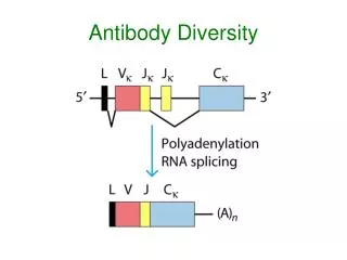
Rearrangement of V, D, and J segments produces a functional heavy-chain gene. Separate gene segments encode the constant domains of the various Immunoglobulin isotypes
Gene rearrangement and the synthesis of cell-surface IgM in B cells SUMMARY
Each B-cell produces immunoglobulin of a single specificity Both the heavy and the light chain coding sequences are present twice in the germline.. (maternal and paternal chromosome) Yet , the B-cell receptor (BCR) on each B-cell is mono-specific The B-cell produces monospecific antibodies This is important for the efficiency of clonal selection and to ensure specificity of the immune response. The process that ensures monospecificity is called: Allelic exclusion
Y a B a/a Y b B b/b Y a B Y Y Y b a B B b a/b A clever genetic model provides evidence for allelic exclusion ALLOTYPE- a polymorphism in the Heavy chain C region of Ig Allotypes can be identified by staining B cell surface Ig with antibodies AND
S. typhi S. typhi Allelic exclusion is needed for efficient clonal selection Antibody All daughter cells must express the same Ig specificity otherwise the efficiency of the response would be compromised Suppression of H chain gene rearrangement helps to prevent the emergence of new daughter specificities during proliferation after clonal selection
Y Y Y Y Y B Self antigen B Y Y Y Y S. aureus S. aureus Y Y Y Y Y Y Y Y Y Y Y Y Anti S. aureus Antibodies Anti S. aureus Antibodies Anti self Abs Y Y Y Y Y Y Y Y Allelic exclusion prevents unwanted responses One Ag receptor per cell IF there were two Ag receptors per cell Suppression of H chain gene rearrangement ensures only one specificty of Ab expressed per cell. Prevents induction of unwanted responses by pathogens
One specificity of Agreceptor per cell IF there were two specificitiesof Ag receptor per cell Y Y Y S. aureus Y B B Y B B B B Y OR Y Y B B Deletion Anergy Allelic exclusion is needed to prevent holes in the repertoire Anti-brain Ig Anti-self Ig AND anti-S. Aureus Ig Exclusion of anti-brain B cells i.e. self tolerance anti S.Aureus B cells will be excluded leaving a “hole in the repertoire” BUT
Allelic exclusion helps diagnose and monitor lymphoma: Due to clonal expansion of a single cell that contains a unique rearrangement the amount of cancer cells in blood or in bone marrow can be determined Can be used to monitor residual tumor cells upon treatment
ChildhoodAcutelymphoblasticleukemia (ALL) is a heterogenousdisease Prognosisvaries Personalizedtreatment is required Minimal residual disease (MRD) Response to therapy Early detection of relapse Bone marryw transplantation
Metods for detection of MRD - nyomonkövetésére használt módszerek • Flow cytometry: • Detection of the aberrynt protein • Kvantitative method, but labour intensive, and less sensitive • Real-time kvantitative PCR • Qquantitative, specificity and superior sensitivity, • Requires minimal amounts os marerial for the test
Diagnosis and monitoring of the treatment of childhood acute lymphoblastic leukemia (C-ALL) The sequence of the framework regions are well conserved. Step 1: design primers that amplify these regions together with the hypervariable regions Step 2: sequence the PCR fragments to obtain spec. Sequence info on the actual tumor (monoclonal, majority of amplification product is tumor-derived Step 3: based on the sequence info, design tumor-specific PCR primers Step 4: quantitate gene expression by Q-PCR. Follow success of therapy and detect minimal residual disease much earlier then by flow cytometry
Result of IMGT junction analysis (http//imgt.cines.fr): IGH rearrangement of patient Q015 I. Identification of monoclonal, i.e. blast-specific gene rearrangements (IGH, TCR, TCR and IGK-KDE) II. Sequencing amplified DNA An algorythm is available for the identification of unique and conserved portions of the sequence ImMunoGeneTics information system (IMGT)
BIOMED2 Concerted Action: IgH, TCRg, TCRd, TCRb, IgK Patient specific forward primer designed by us Consensus TaqMan probe and reverse primer V N1 D N2 J Design patient-specific primers (red) and use them together with consensus primers