Download
1 / 31
310 likes | 432 Views
Clinical Disorders and Diseases of the Skeletal System. Cleft Lip/Palate. Facial and oral malformations that occur very early in pregnancy Results when there is not enough tissue in the mouth or lip area, and the tissue that is available does not join together properly

E N D
Cleft Lip/Palate • Facial and oral malformations that occur very early in pregnancy • Results when there is not enough tissue in the mouth or lip area, and the tissue that is available does not join together properly • Cleft lip – split or separation of the two sides of the upper lip • Cleft palate – split or opening in the roof of the mouth (hard or soft palate) • 1 in 700 babies; 4th most common birth defect in the US
Cleft lip/palate cont. • The cause is unknown • May be linked to genetic and environmental factors (drugs, exposure to viruses or chemicals) • Eating, speech, and dental problems could result • Often requires multiple surgeries to treat
Vertebral Column: Curvatures Scoliosis: abnormal lateral curvature of the spine (occurs most often in the thoracic region) • Caused by a bone abnormality present at birth, abnormal muscles or nerves, trauma, or genetic • 2-3% of Americans at age 16 (girls are more prone to developing the condition) • Diagnosed by screening exams, bone exam, and X-ray • Treatments include braces or surgery (spinal fusion)
Osteomalacia • Softening of the bones due to a lack of Vitamin D or a problem with the body’s ability to break down and use Vitamin D • Rickets - Children's form of osteomalacia • Causes – not enough Vitamin D; not enough exposure to sunlight or malabsortption of Vitamin D by the intestines • Symptoms - bone weakness, fractures that occur without real injury, and numbness • Treatments – Vitamin D, calcium, and phosphorus supplements
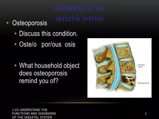
Osteoporosis • Bone loss outpaces bone regeneration • Bones weaken and lose mass • Bones become brittle and fractures occur more often • Found most often in women • Treatment may include; medication, diet changes, exercise
Osteoarthritis • Degenerative joint disease • Most common type of arthritis (21 million) • Breakdown of cartilage in joints • Mostly occurs in the weight bearing joints, but it can occur anywhere • Causes cartilage to become stiff and lose its elasticity • As cartilage deteriorates, tendons and ligaments stretch, causing pain
Osteoarthritis • Symptoms: • Joint aching and soreness • Pain after overuse or long periods of inactivity • Joint swelling • Fluid accumulation • Treatment: medication, physical therapy, surgery
Knee Replacement surgery • Generally reserved for people over the age of 50 with severe osteoarthritis • Helps relieve pain & restore function in severely diseased knee joints • During surgery; a surgeon cuts away damaged bone and cartilage from your femur, patella, and tibia and replaces it with an artificial joint made of metal alloys, high-grade plastics, and polymers
Fractures • A crack or break in a bone • Despite its mineral strength, bone can crack or even break if subjected to extreme loads, sudden impacts, or stresses from unusual directions
Types of Fractures • Named according to their external appearance, their location, and the nature of the crack or break in the bone. • Two general categories: • Closed (simple) – fracture is internal • Open (compound) – fracture projects through the skin
Spiral fractures Figure 6–16 (4 of 9)
Greenstick fracture Figure 6–16 (7 of 9)
Compression fractures Figure 6–16 (9 of 9)
Treatment of a Fracture • Initial treatment for fractures of arms, legs, hands, and feet include splinting the extremity in the position it is found, elevation, and ice. • Edema (or swelling) What does this have to do with splinting and casting? • Closed Reduction – manual realignment • Open Reduction – surgically realignment
Steps in the Repair of a Fracture Step 1 – • Immediately after the fracture, extensive bleeding occurs (blood vessels are broken). • A large blood clot, or fracture hematoma, soon closes off the injured vessels and leaves a fibrous meshwork in the damaged area. • The disruption of the circulation kills osteocytes (mature bone cells) around the fracture. • Dead bone soon extends along the shaft.
Steps in the Repair of a Fracture Step 2 – • The cells of the endosteum (cellular layer) and periosteum undergo cell division and the daughter cells migrate into the fracture zone. • An external callus (hard skin) forms and encircles fracture • An internal callus organizes within the cavity and between the broken ends of the shaft • The broken ends have been temporarily stabilized
Steps in the Repair of a Fracture Step 3 – • Osteoblasts (bone building cells) replace the central cartilage of the external callus with spongy bone • Calluses form a brace at the fracture site • Spongy bone now unites the broken ends • Fragments of dead bone are removed and replaced • If the fracture required a cast, it can be removed at this stage
Steps in the Repair of a Fracture Step 4 – • Osteoclasts (remove and recycle bone matrix) and osteoblasts continue to remodel the region of the fracture (4 months to 1 year) • When remodeling is complete, the bone of the calluses is gone and only living compact bone remains. • The bone could be slightly thicker and stronger than normal at the fracture site
Casts • Holds a broken bone in place as it heals • Help to prevent or decrease muscle contractions • Provide immobilization (the joints above and below the area) • Casts are made of plaster and fiberglass • Typically worn for 6-8 weeks
Dislocation • Separation of two bones where they meet at a joint (no longer in normal position) • Caused by a sudden impact to the joint • May be hard to tell a dislocated bone from a broken bone • Generally take 3-6 weeks to heal • Possible ligament damage can occur