Download
1 / 29
310 likes | 590 Views
Gout. Dr. Müge Bıçakçıgil Kalaycı. GOUT. Common medical problem, Affects at least 1 percent of men in Western countries, Male: female ratio 7:1 to 9:1. Primary causes Idiopathic HGPRT deficiency Increased PRPP activity. Secondary causes Excessive dietary purine intake

E N D
Gout Dr. Müge Bıçakçıgil Kalaycı
GOUT • Common medical problem, • Affects at least 1 percent of men in Western countries, • Male: female ratio 7:1 to 9:1.
Primary causes Idiopathic HGPRT deficiency Increased PRPP activity Secondary causes Excessive dietary purine intake Increased nucleotide turnover Myeloproliferative disease Lymphoproliferative disease Hemolytic anemia Psoriasis Accelerated ATP degradation Hereditary fructose intolerance Glycogen storage disease Severe muscle exertion Ethanol abuse Uric Acid Overproduction
Renal insufficiency Inhibition of tubular urate secretion: Keto- and lactoacidosis Enhanced tubular urate reabsorption: Diuretics, Insulin resistance, Dehydration Undefined mechanism: Hypertension, Hyperparathyroidis Low-dose salicylates, Pyrazinamide, Ethambutol Lead nephropathy Uric Acid underexcretion
GOUT Clinical & Laboratory Features
Stages - Asymptomatic Hyperuricemia • In physiological terms any level above 6.8 mg/dl is hyperuricemia, since it exceeds the soluble concentration of MSU in body fluids. • Vast majority of people with hyperuricemia will never develop symptoms.
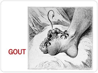
Stages -Acute intermittent gout • Characteristic gout attack: • rapid development of warmth, swelling, erythema and pain in the affected joint. • The initial attack • is monoarticular and • in 50% of cases involves the 1st metatarsal joint, which will finally be affected in 90% of patients.
Stages -Acute intermittent gout • Other joints: MT, ankle, heels and knees. • Systemic symptoms: Fever, chills and malaise. • Early in the disease the episodes are infrequent • Between the attacks the previously affected joints are free of pain, • despite this, MSU crystals can be identified in the synovial fluid.
Stages – Chronic Tophaceous Gout • Usually develops after 10 years of acute intermittent gout. • In this stage the affected joints become persistently uncomfortable and swollen. • The intensity of these symptoms is much less than the acute attacks.
Stages – Chronic Tophaceous Gout • Characterized by: • tophi formation and • polyarticular involvement, including the small joints of the hands . • Subcutaneous gouty tophi can be found in the fingers, wrists, ears, knees, olecranon bursa and pressure points
Provocative factors • The degree of decrease or increase in the concentration of synovial-fluid urate is more related to acute attack than the degree of hyperuricemia . • Traumais frequently reported as an initiating event for an acute gouty attack: • the attack occurs when the joint is allowed to rest, • there is arapid efflux of water from the joint fluid and the result is sudden increase in urate concentration.
Provocative factors • Alcohol ingestion: • By accelerating the breakdown of intracellular ATP • Alcohol contains large quantities of guanosine. • Drugs: • thiazides, • low dose aspirin.
Clinical association – Renal involvement • Chronic urate nephropathy: • Deposition of MSU in the renal medulla and pyramids, • Associated with mild microalbuminuria. • Acute uric acid nephropathy: • ARF caused by hyperuricemia • in tumor lysis syndrome or post chemotherapy. • Uric acid renal stones: • 10-25% of all people with gout, • the incidence correlate with the serum urate levels.
Radiological features • In early stages : • Soft tissue swelling around the affected joints • Preserved joint space • Later: • Bony erosions that are both atrophic and hypertrophic, • Erosions with overhanging edges
Laboratory Features and Diagnosis • Uric acid level in serum is of limited value in establishing the diagnosis: • The majority of hyperuricemic subjects will not develop gout. • Normal level of uric acid during gouty attack is frequent.
Diagnosis • Definitive diagnosis is possible only by aspiration and inspection of the synovial fluid or tophaceous material. • Crystals are needle or rod-shaped. • On compensated polarized microscopy, they appear as a bright, birefringent crystals (usually intracellular) that are yellow when parallel to the axis of slow vibration.
Treatment • The management of gout involves • treating acute arthritic inflammation and urolithiasis • lowering urate levels with the goal of preventing recurrent disease and progression.
Treatment of Acute Gouty Arthritis • NSAIDs are considered first-line therapy. • Selective Cox-2 inhibitors are an alternative in patients with GI contraindications. • Corticosteroids or subcutaneous injections of corticotropin are additional alternatives. • Because colchicine adverse effects can be serious, IV colchicine should not be used.
Long-Term or Prophylactic Therapy • NSAIDs and colchicine are frequently used as prophylaxis against recurrent acute gout, since such episodes are common during the initiation of uric acid–lowering treatment. • Allopurinol and • Probenecid - apotent uricosuric agents equally acceptable as first-line drug.
CPPD Crystals Deposition Disease(PSEUDOGUT- CHONDROCALCİNOSİS) Can cause monoarthritis clinically indistinguishable from gout – Pseudogout. Rheumatic manifestation of calcium pyrophosphate crystal deposition in articular cartilage (chondrocalcinosis), synovium, and periarticular ligaments and tendons
Pseudogout is most common in the knee (50%) but often also the wrist, elbow, shoulder,ankle and hand. • Identification of crystals is the only means of positive diagnosis. • Increasing age is a predisposing factor; more than 80% of patients are over 60 years old. • Peak age 65-75.
Affects one or a few joints; typically those that are weight bearing or have suffered previous injury • Slowly progressive with occasional aexacerbations , which are usualy not associated with effusion, warmth, and redness • No rest pain (unless disease is end stage ) symptoms worse with or after sustained activity • Joints show bony swelling, crepitus, and restricted movement
Ca pyrophosphate (pseudogout) Rod or rhomboid-shaped Weakly positive birefringent