Download
1 / 60
640 likes | 905 Views
High grade gliomas diagnosis management and controversies. introduction. High-grade gliomas (HGGs) represent a heterogenous group of tumors and account for most primary brain tumors. Despite aggressive therapies, they are invariably associated with poor patient outcome. Include -

E N D
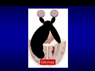
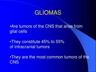
introduction • High-grade gliomas (HGGs) represent a heterogenous group of tumors and account for most primary brain tumors. • Despite aggressive therapies, they are invariably associated with poor patient outcome. • Include - 1) anaplastic (World Health Organization [WHO] grade III) histologies of • astrocytomas, • oligodendrogliomas • ependymomas 2) WHO grade IV glioblastomamultiforme (GBM).
Glioma Grading AstrocytomaOligodendroglioma mixed Median survival (range) • Grade II: 5 yr (3-10y) • Nuclear atypia • Grade III: 3 yr • Nuclear atypia + mitosis • Grade IV: 1 yr • Nuclear atypia + mitosis + either endothelial proliferation and/or necrosis • Grade II: 15 yr (8-20y) • Grade III: 3 yr • The World Health Organization (WHO) scheme is based on the appearance of certain characteristics: atypia, mitoses, endothelial proliferation, and necrosis
Epidemiology • Annual incidence approximately 5 cases per 100,000. • Glioblastomas account for approximately 60 to 70% of malignant gliomas, - anaplastic astrocytomas for 10 to 15%, - anaplastic oligodendrogliomas and anaplasticoligoastrocytomas for 10%; - anaplastic ependymomas and anaplastic gangliogliomas account for the rest. • 2008 statistical report: primary brain tumors in the United States, 1998-2002. Central Brain Tumor Registry of the United States, 2000-2004.
india • Among series of 500 patients with glioma(AIIMS) - 222 (44 %)patient had high grade glioma. - 73.6 % were male. • Banerji AK epidemiology of CNS tumours(1974-78) neuro oncology 1981 p 43 Tandon PN supratentorial gliomas neurolindia, 42;131,1994
The median age of patients at the time of diagnosis is 64 years in the case of glioblastomas 45 years in the case of anaplastic gliomas. • Epidemiology of brain tumors. NeurolClin 2007;25:867-90.
MOLECULAR PATHOGENESIS • Glioblastomas - two main subtypes • on the basis of biologic and genetic differences. Primary Glioblastomas - occur in patients older than 50 years - characterized by EGFR amplification and mutations, loss of heterozygosity of chr 10q and p16 deletion. • Phillips HS, Kharbanda S, Chen R, et al. Molecular subclasses of high-grade glioma predict prognosis, delineate a pattern of disease progression, and resemble stages in neurogenesis. Cancer Cell 2006;9
Secondary glioblastomas • younger patients as low-grade or anaplasticastrocytomas transform over a period of several years into glioblastoma. • less common than primary glioblastomas, • characterized by mutations in the p53 tumor suppressor gene, over expression of the platelet derived growth factor receptor (PDGFR), abnormalities in the p16 and retinoblastoma (Rb) pathways, and loss of heterozygosity of chromosome 10q. • Furnari FB, Fenton T, Bachoo RM, et al. Malignant astrocytic glioma: genetics, biology, and paths to treatment. Genes Dev 2007;21:2683-710.
Despite their genetic differences primary and secondary glioblastomas are morphologically indistinguishable and respond similarly to conventional therapy, • but they may respond differently to targeted molecular therapies. High-grade oligodendrogliomas Characterized by the loss of chromosomes 1p and 19q (in 50 to 90% of patients).
Deregulated Growth Factor Signaling • Most common defects in growth-factor signaling involve EGFR and PDGFR. • EGFR gene amplification is one of the most common genetic events in glioblastomas. • The most common EGFR mutant is the constitutively active EGFRvIII. • Seen almost exclusively in primary glioblastomas and is seen in approximately 40 to 50% .
diagnosis Clinical Radiographical Pathologic
Clinical Presentation(Varies depending upon size and location of tumor) Most common symptoms: Headache (80%) Seizure (30%) Focal neurologic deficits Change in mental status Time from initial symptoms to diagnosis usually < 6 months (70% of patients)
radiology • CT HEAD • Features - nonspecific. • Overlap of imaging features between low- and high-grade gliomas (HGG).
“Density” • Variable • Usually isodense to moderately hyperdense to gray matter (the isocenter of the mass may appear denser because of the surrounding vasogenic edema). • Small or atypically hypo dense mass may not be visible on no enhanced CT study, masked by the presence of the surrounding, low-density, vasogenic edema- ?infarct
Hemorrhage • depicted as an area of high density • commonly seen in advanced grade gliomas • (World Health Organization [WHO] grade III– IV), particularly glioblastomas • not a pathognomonic feature;
Calcifications • Low-grade gliomas are more likely to be calcified • small percentage of HGG also shows calcifications • Prognostic significance of CT contrast enhancement within histological subgroups of intracranial glioma. J Neurooncol 1998;40:161–170.
Vasogenic Edema • indirect indicator of the aggressive growth of a brain tumor • responsible for the secondary mass effect. • Focal region of vasogenic edema may be the only finding on unenhanced CT in cases of HGG or intracranial metastases. White matter edema produced by HGG, particularly GBM, can be quite extensive and actually represents a tumor plus edema • Earnest F, Kelly PJ, Scheithauer BW, et al. Cerebral astrocytomas: histopathologic correlation of MR and CT contrast enhancement with stereotactic biopsy. Radiology 1988;166:823–827.
Contrast Enhancement • Contrast enhancement high sensitivity and specificity for HGG (about 87 and 79%, respectively) . • 20% of low-grade gliomas . • Likewise, a substantial number of nonenhancing brain tumors can turn out to be HGG, particularly anaplastic astrocytomas . • Enhancement on CT imaging- negative prognostic factor, irrespective of the glioma grade. • contrast enhanced area (CEA) of >4 cm and heterogenous enhancement as negative prognostic factors . • CEA <4cm and homogenous enhancement indicates better prognosis. Yamada S, Takai Y, Nemoto K, et al. Prognostic significance of CT scan in malignant glioma. Tohoku J ExpMed 1993;170:35–43.
Anaplastic astrocytomas • Because of their infiltrative growth, indistinct margins - ill-defined, inhomogeneous lesions on CT. • High cellularity- the attenuation usually ranges from intermediate to high in relation to the normal brain. • Cystic change or necrosis and hemorrhage are uncommon. • AA more homogenous compared with GBM. • Calcification is rare. • Post-contrast CT studies usually show limited to moderate patchy/ heterogenous enhancement .
GlioblastomaMultiforme • NCCT - mixed density complex mass with disproportionate surrounding vasogenic edema, usually involves corpus callosum . • Marked heterogeneity - necrosis and hemorrhage. • Central necrosis is an imaging hallmark of GBM. • Hemorrhage is seen in about 20% of GBM. • Vasogenic edema is a prominent imaging feature of GBM and commonly extends along the white matter tracts. • GBM typically shows heterogeneous rim enhancement with irregular thick and nodular margins .
Anaplastic Oligodendroglioma /Oligoastrocytoma • Involve cortex and sub cortical white matter. • Typical finding is a well defined mixed density superficial frontal lobe mass with calcifications. • Nodular, clumped, or even gyriform. • Involved cortex is expanded, cystic degeneration is common, and hemorrhage or necrosis may also be seen. • Lesions may expand, remodel, or erode the calvarium.
Gliosarcoma • Subtype of GBM in which a sarcomatous component is there. • Typically present along the periphery, abutting the dura. • Sharply defined, round or lobulated, hyperdense solid mass • Homogeneous contrast enhancement and peri-tumoral edema. • Tendency to invade adjacent dural reflections . • Mimics meningioma; both in imaging and surgery. • More heterogenous, have a smaller dural based attachment • Significant amount of parenchymal edema.
Advantages OF CT • Superior in detecting calcification, hemorrhage and in evaluating bone changes related to tumors. • Pacemakers or metallic devices as well as critically ill or unstable patient. • Assessment of immediate post-operative complications.
Magnetic Resonance Imaging • Infiltrative parenchymal masses. • Hyperintense on FLAIR and T2-weighted images. • Hypointense on unenhanced T1-weighted images. • May or may not extend into the corpus callosum. • Surrounded by extensive vasogenic edema. • Prominently enhance following gadolinium administration.
Treatment • Surgery • Radiation • Chemotherapy
Summary of Current Treatments for Malignant Gliomas. • Type of Tumor Therapy • Newly diagnosed tumors Glioblastomas (WHO grade IV). Maximal surgical resection, plus radiotherapy, plus concomitant and adjuvant TMZ or carmustine wafers (Gliadel). • Anaplastic astrocytomas (WHO grade III) Maximal surgical resection, with the following options after surgery (no accepted standard treatment): radiotherapy, plus concomitant and adjuvant TMZ or adjuvant TMZ alone. Data are from Sathornsumetee et al.,3 Furnari et al.,18 Chi and Wen,20 and Sathornsumetee et al.21 • PCV denotes procarbazine, lomustine (CCNU), and vincristine, and TMZ temozolomide • Sathornsumetee S, Rich JN, Reardon DA. Diagnosis and treatment of highgradeastrocytoma. NeurolClin 2007;25: • 1111-39.
Anaplastic oligodendrogliomas and anaplastic oligoastrocytomas (WHO grade III) Maximal surgical resection, with the following options after surgery (no accepted standard treatment): radiotherapy alone, TMZ or PCV with or without radiotherapy afterward, radiotherapy plus concomitant and adjuvant TMZ, or radiotherapy plus adjuvant TMZ. • Recurrent tumors Reoperation in selected patients, carmustine wafers (Gliadel), conventional chemotherapy (e.g., lomustine, carmustine, PCV, carboplatin, irinotecan,etoposide), bevacizumab plus irinotecan, experimental therapies.
Surgical Resection • Incurable secondary to the infiltrative nature • Rationale behind resection: -to obtain definitive histologic diagnosis . -to palliate symptoms from local tumor effect. -to potentially provide better tumor control. with radiation/chemotherapy . -to provide tissue for molecular/genetic analysis. prognostication and research. -to provide improved survival.
Controversy exists behind the correlation between the extend of resection and survival. • Goal is to remove as much tumor without causing neurologic dysfunction. • Those that cannot be removed will need a stereotactic/open needle biopsy. • Recently that there is class I evidence to support this long held belief that aggressive, safe surgical resection is an important favorable prognostic factor, even in the elderly . • Vuorinen V, Hinkka S, Farkkila M, Jaaskelainen J, Debulking or biopsy of malignant glioma in elderly people– randomized study. ActaNeurochir (Wien) 2003;145(1):5–10
STEREOTACTIC BRAIN BIOPSY • patients who have inoperable tumors • that a located in critical areas • confirm histological diagnosis . r/o other pathology like PCNSL • additional molecular information Determining the status of chromosomes 1p and 19q in anaplastic oligodendroglioma
Biopsy versus resection in the management of malignant gliomas: a systematic review and meta-analysis A review • J Neurosurg 112:1020–1032, 2010 • Abraham Tsitlakidis, M.D., M.Sc.,1 Nicolas Foroglou, M.D., Ph.D.,1 • Christos A. Venetis, M.D., M.Sc.,2 Ioann is Pat salas, M.D., Ph.D.,1 • Athanasios Hat zisotiriou, M.D., Ph.D.,3 an d PanagiotisSelviaridis, M.D., Ph.D.1 • 1First Department of Neurosurgery, AHEPA University Hospital; 2Unit for Human Reproduction, First Department of Obstetrics and Gynaecology, Papageorgiou General Hospital, Aristotle University of Thessaloniki; and 3Agios Loukas Clinic, Thessaloniki, Greece
One randomized controlled trial and 4 retrospective studies (involving a total of 1111 patients) were found eligible for this systematic review. • A meta-analysis demonstrated a significant increase in overall survival in the patients treated with resection instead of biopsy ( p < 0.0001). However, there did not seem to be any significant difference in progression-free survival between the 2 groups. • Well designed prospective studies are needed for more solid conclusions to be drawn.
Radiation Therapy • Mainstay of treatment increases survival among patients with glioblastomas from a range of 3 to 4 months to a range of 7 to 12 months. Conventional radiotherapy • 60 Gy of partial-field external- beam irradiation delivered 5 days per week in fractions of 1.8 to 2.0 Gy. • Walker MD, Alexander E Jr, Hunt WE, et al. Evaluation of BCNU and/or radiotherapy in the treatment of anaplastic gliomas: a cooperative clinical trial. J Neurosurg 1978;49:333-43. • Stupp R, Mason WP, van den Bent MJ, et al. Radiotherapy plus concomitant and adjuvant temozolomide for glioblastoma. N Engl J Med 2005;352:987-96.
Strategies to increase the radiation dose to the tumor with the use of brachytherapy . • Stereotactic radiosurgery have failed to improve survival. • Souhami L, Seiferheld W, Brachman D, et al. Randomized comparison of stereotactic radiosurgery followed by conventional radiotherapy with carmustine to conventional radiotherapy with carmustine for patients with glioblastomamultiforme: report of Radiation Therapy Oncology Group 93-05 protocol. Int J RadiatOncolBiol Phys 2004;60:853-60.
Patients older than 70 years of age have a worse prognosis. • Radiotherapy produces a modest benefit in median survival (29.1 weeks) as compared with supportive care (16.9 weeks). • Older patients often tolerate radiotherapy poorly. • An abbreviated course of radiotherapy (40 Gy in 15 fractions over a period of 3 weeks) or chemotherapy with temozolomide (an oral alkylating agent with good penetration of the blood–brain barrier) alone. Outcomes with these approaches are similar to the outcomes with conventional radiotherapy regimens. Keime-Guibert F, Chinot O, Taillandier L, et al. Radiotherapy for glioblastoma in the elderly. N Engl J Med 2007;
chemotherapy • Chemotherapy is assuming an increasingly important role in the treatment. • Early studies of adjuvant chemotherapy for malignant gliomas with the use nitrosoureas failed to show a benefit. • Randomized trial of procarbazine, lomustine, and vincristine in the adjuvant treatment of high-grade astrocytoma: a Medical Research Council trial. J ClinOncol 2001;19:509-18. • Alexander E Jr, Hunt WE,et al. Evaluation of BCNU and/or radiotherapy in the treatment of anaplastic gliomas: a cooperative clinical trial. J Neurosurg1978;49:333-43.
Two metaanalyses have suggested that adjuvant chemotherapy results in a modest increase in survival (a 6 to 10% increase in the 1-year survival rate). • Fine HA, Dear KB, Loeffler JS, Black PM, Canellos GP. Meta-analysis of radiation therapy with and without adjuvant chemotherapy for malignant gliomas in adults. Cancer 1993;71:2585-97. • Stewart LA. Chemotherapy in adult high-grade glioma: a systematic review and meta-analysis of individual patient data from 12 randomised trials. Lancet 2002;359:1011-8.
The European Organisation for Research and Treatment of Cancer (EORTC) and the National Cancer Institute of Canada (NCIC) conducted a phase III trial comparing radiotherapy alone (60 Gy over a period of 6 weeks) with radiotherapy and concomitant treatment with temozolomide (75 mg per square meter of body-surface area per day for 6 weeks), followed by adjuvant temozolomide therapy (150 to 200 mg per square meter per day for 5 days every 28 days for 6 cycles), in patients with newly diagnosed glioblastomas.
The combination of radiotherapy and temozolomide had an acceptable side-effect profile and, as compared with radiotherapy alone increased the median survival (14.6 months vs. 12.1 months, P<0.001). • In addition, the survival rate at 2 years among the patients who received radiotherapy and temozolomide was significantly greater than the rate among the patients who received radiotherapy alone (26.5% vs. 10.4%), • . Stupp R, Mason WP, van den Bent MJ,et al. Radiotherapy plus concomitant and adjuvant temozolomide for glioblastoma.NEngl J Med 2005;352:987-96.
mgmt • MGMT is an important repair enzyme that contributes to resistance to temozolomide. • Patients with glioblastoma and MGMT promoter methylation (45% of the total) who were treated with temozolomide had a median survival of 21.7 months and a 2-year survival rate of 46%. • In contrast, patients without MGMT promoter methylation who were treated with temozolomide had a significantly shorter median survival of only 12.7 months and a 2-year survival rate of 13.8%. Hegi ME, Diserens AC, Gorlia T, et al. MGMT gene silencing and benefit from temozolomide in glioblastoma. N Engl J Med 2005;352:997-1003.
Currently, temozolomide is used in the treatment of glioblastomas regardless of MGMT promoter methylation status. However, if the importance of MGMT promoter methylationis confirmed by the results of an ongoing study by the Radiation Therapy Oncology Group (RTOG 0525), patients with unfavorable MGMT methylation status may be selected for other treatments in future investigations.
implantation of biodegradable polymers containing carmustine (Gliadel Wafers, MGI Pharma) into the tumor bed after resection of the tumor. • Aim of treatment with these polymers is to kill residual tumor cells. • In a randomized, placebo-controlled trial that investigated the use of these polymers in patients with newly diagnosed malignant gliomas, median survival increased from 11.6 months to 13.9 months (P = 0.03). This survival advantage was maintained at 2 and 3 years. • Westphal M, Ram Z, Riddle V, Hilt 78. D, Bortey E. Gliadel wafer in initial surgery for malignant glioma: long-term followup of a multicenter controlled trial. ActaNeurochir (Wien) 2006;148:269-75.
Therapy for Anaplastic Gliomas • Anaplastic astrocytomas are treated with radiotherapy and either concurrent and adjuvant temozolomide (as for glioblastomas) or adjuvant temozolomide alone.
Anaplastic oligodendrogliomas and anaplastic oligoastrocytomas • Important subgroup of malignant gliomas that are generally more responsive to therapy than are pure astrocytic tumors. • Tumors in patients with the 1p and 19q codeletion are particularly sensitive to chemotherapy with PCV — procarbazine, lomustine (CCNU), and vincristine — with response rates of up to 100%, as compared with response rates of 23 to 31% among patients without the deletion of chromosomes 1p and 19q. • van den Bent MJ. Anaplastic oligodendroglioma and oligoastrocytoma. NeurolClin 2007;25:1089-109.
Two large phase III studies of PCV chemotherapy with radiotherapy, as compared with radiotherapy alone, in patients with newly diagnosed anaplasticoligodendrogliomas or anaplastic oligoastrocytomas, have been reported. • In both studies, the addition of chemotherapy to radiotherapy increased the time to tumor progression by 10 to 12 month but did not improve overall survival (median, 3.4 and 4.9 years). • Cairncross G, Berkey B, Shaw E, et al. Phase III trial of chemotherapy plus radiotherapy compared with radiotherapy alone for pure and mixed anaplastic oligodendroglioma: Intergroup Radiation Therapy Oncology Group Trial 9402. J ClinOncol 2006;24:2707-14. van den Bent MJ, Carpentier AF, BrandesAA, et al. Adjuvant procarbazine, lomustine, and vincristine improves progression-free survival but not overall survival in newly diagnosed anaplastic oligodendrogliomas and oligoastrocytomas: a randomized European Organisation for Research and Treatment of Cancer phase III trial. J ClinOncol 2006;24:2715-22.
Management of malignant glioma: progress in multimodal approaches
Therapy for Recurrent Malignant Gliomas • In patients without medical contraindications Surgery - can confirm tumor recurrence, - reduce intracranial pressure, improve neurological status, and possibly improve efficacy of adjunctive therapy. Stereotatically guided biopsy procedures allow for - the sampling of small, inaccessible, or even multiple lesions with minimal patient morbidity and mortality rates (estimated to be 2–5% and , 1% respectively). Treatment decisions for patients whose imaging studies fail to differentiate between radiation necrosis and tumor recurrence.
In addition to decreasing mass effect, repeated craniotomy allows for the potential in situ delivery of chemotherapy or brachytherapy. • Randomized study Brem, et al evaluating the efficacy of BCNU implantation during repeated resection compared with placebo, reported a 50% improvement in survival at 6 months (56% with BCNU compared with 36% with placebo). • Preoperative performance status and age were significant prognostic factors. • Resection should be seriously considered in those with a high KPS score (70) and whose lesions are in a favorable location. Brem H, Piantadosi S, Burger PC, et al: Placebo-controlled trial of safety and efficacy of intraoperative controlled delivery by biodegradable polymers of chemotherapy for recurrent gliomas. The Polymer-brain Tumor Treatment Group. Lancet 345: 1008–1012, 1995
Rostomily et al., reported a prolonged PFS of 7 weeks in patients undergoing combined chemotherapy plus repeated resection compared with patients receiving chemotherapy alone (21 weeks compared with 14 weeks). • Overall survival rate among this cohort of 51 patients was equivocal. Rostomily RC, Spence AM, Duong D, et al: Multimodality management of recurrent adult malignant gliomas: results of a phase II multiagent chemotherapy study and analysis of cytoreductive surgery. Neurosurgery 35:378–388, 1994
chemotherapy • Chemotherapy is the most common treatment option for recurrent malignant gliomas. • Traditionally reserved for salvage treatment of recurrent GBM, • Administered alone or as a supplement to cytoreductive surgery. • Chemotherapeutic agents such as TMZ, carboplatin, procarbazine, and imatinibmesylate are currently being examined for their potential in the palliative treatment of recurrent GBMs.