Download
1 / 37
651 likes | 1.93k Views
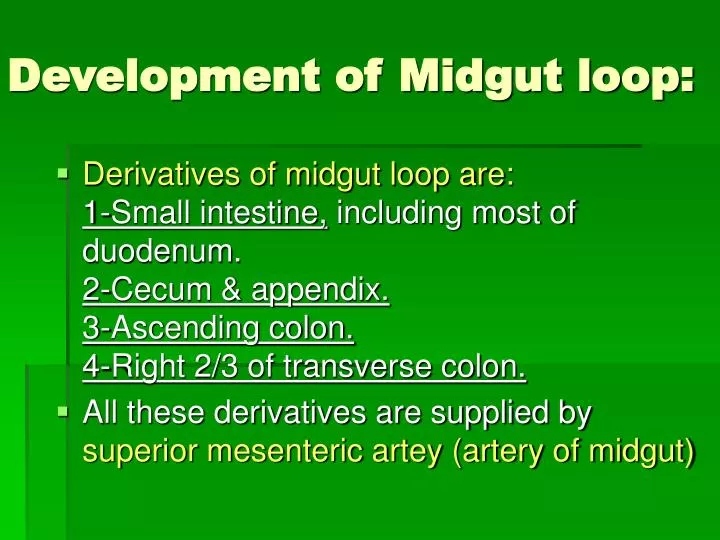
Development of Midgut loop:. Derivatives of midgut loop are: 1-Small intestine, including most of duodenum. 2-Cecum & appendix. 3-Ascending colon. 4-Right 2/3 of transverse colon.

E N D
Development of Midgut loop: • Derivatives of midgut loop are:1-Small intestine, including most of duodenum. 2-Cecum & appendix.3-Ascending colon.4-Right 2/3 of transverse colon. • All these derivatives are supplied by superior mesenteric artey (artery of midgut)
Development of midgut loop • At the biginning of 6th week, the midgut elongates to form a venteral U-shaped midgut loop projecting into extra-embryonic coelom of proximal part of umbilical cord …this called physiological umbilical herniation. • At this stage the arrow indicates communication of intraembryonic coelom (peritoneal cavity) with extraembryonic coelom. • At this stage ,the abdominalcavity is temporarily too small in comparison to relatively massive liver & kidnes to contain the developing rapidly growing intestines.
Development of midgut loop • The midgut loop joined with Yolk sac through yolk stalk or vitelline duct until 10th week. • Midgut loop consists of 2 limbs, cranial limb & caudal limb.(A) • Cranial limb grows rapidly forming the small intestines.(B) • Caudal limb gives rise to cecal diverticulum, the primordium of cecum & appendix. ( C )
Rotation of midgut loop • While midgut loop is in the umbilical cord, it rotates 90 degrees counterclockwise around axis of superior mesenteric artery. (A),(A1) • This brings the cranial limb tothe right and caudal limb to left.(B), (B1).
Return of midgut to abdomen (reduction of physiological hernia) : • During 10th week the small intestine (formed of cranial limb) returns first to abdomen due to enlagement of abdominal cavity ….this is called reduction of physio-logical midgut hernia.(C&D) • large intestine (formed from caudal limb) undergoes a further 180 degree counterclockwise rotation to occupy right side of abdomen. (D),(D1) • Ascending colon becomes recognizable as posterior abdominal wall elongates. (E)
Fixation of various parts of intestines • At first the dorsal mesentry is in the median plane. As the intestines lenghthen,and obtain their final position, some mesentries are fused with posterior abdominal wall. • Mesentry of ascending& descending colondisappears, so ascending & descending colon becomes fixed to posterior abdominal wall and they become retro-peritoneal. (B) • Transverse mesocolonpersists and fuses with posterior wall of greater omentum and maintains its mobility. (C) A,Venteral view of the intestine Prior to Fixation. B,T.S showing areas of fusion. C, Sagittal S. showing greater omentum over- hanging the transverse colon, arrows indicate areas of subsequent fusion.
Fixation of various parts of intestines • With rotation of stomach, duodenum & pancreas are pressed against posterior abdominal wall by the colon. • Adjacent layers of peritoneum fuse and disappear, so most of duodenum & pancreas become retroperitoneal. Intestines prior to fixation • E, T.S after disappearance of mesentry of ascending & descending colon. • F, sagittal S.showing fusion of layers of greater omentum and fusion ofgreater omentum with mesentery of transverse colon. Intestines after fixation
Development of Cecum & Appendix • At 6th week, cecal diverticulum appears as a swelling on the anti-mesenteric border of caudal limb of midgut loop. (A) • Cecal diverticulum gives rise to cecum & appendix.(B) • Appendix is intially a small diverticulum of the cecum, arising from distal end of cecum.(B) • As ascending colon elongates, appendix elongates and may be retrocecal or retrocolic or pelvic appendix. In 64% of people, it is retrocecally. • A,6 weeks . B, 8 weeks.C,12 weeks • D,at birth …appendix is long and is continuous with apex of cecum. E, After birth (adult appendix) It is short as a result that the appendix enters medial side of cecum.
Congenital Omphalocele • It is a persistence of herniation of abdominal contents into proximal part of umbilical cord due to failure of reduction ofphysiological hernia to abdominal cavity at 10th week. • Herniation of intestines occurs in 1 of 5000 births – herniation of liver & intestines occurs in 1 of 10,000 births. • It is accompanied by small abdominal cavity. • The hernial sac is covered by the epithelium of the umbilical cord, the amnion. • Immediate surgical repair is required.
Umbilical Hernia • The intestines return toabdominal cavity at 10th week, but herniate through an imperfectly closed umbilicus • It is a common type of hernia. • The herniated contents are usually the greater omentum & small intestine. • The hernial sac is covered by subcutaneous tissue & skin. • It protrudes during crying,straining or coughing and can be easily reduced through fibrous ring at umbilicus. • Surgery is performed at age of3-5 years.
Ileal (Meckel) Diverticulum • It is one of the most common anomalies of the digestive tract, present in about 2% of people. • It is a small pouch from the ileum, and may contain small patches of gastric & pancreatictissues.The gastric mucosa often secretes acid producing ulceration & bleeding. • It is the remnant of proximal part nonobliterated part)of yolk stalk (or vitelline duct). • It arises from antimesenteric border of ileum,1/2 meter from ileocecal junction. • It is more common in males. • It is sometimes becomes inflamed and causes symptoms that mimic appendicitis. • It may be connected to the umbilucus by a fibrous cord or Omphalo-enteric fistula (vitello-intestinal duct remains patent and faecal matter is carried through the duct into umbilicus).
Hindgut • Derivatives of hindgut are : • 1-left 1/3 of transverse colon. 2-Descending colon & sigmoid colon. • Part of hindgut dilate to form Cloaca which gives rise to:3-Rectum.4-Superior part of anal canal. 5-Certain urogenital structures (epithelium of urinary bladder & most of urethra) • Inferior mesenteric arteryis the artery ofhindgut.
Partitioning of cloaca into rectum and uro-genital sinus by urorectal septum. • A,C,E cloaca at 4,6 and 7 weeks. • B,D,F cloacal region.note degeneration and disappearance of the postanal or tailgut (B) as the rectum forms from dorsal part of cloaca. • B1,D1,F1, T.S.of cloaca.
Development of Rectum & Anal canal • Cloaca, the expanded terminal part of hindgut, receives allantois ventrally. • cloacal membrane, is composed of endoderm ofcloaca + ectoderm of proctodeum (or anal pit). • Cloaca is divided into dorsal & ventral parts by a mesenchyme-urorectalseptum- between allantois and hindgut, producing infoldings of lateral walls of cloaca. • These folds fuse forming a partition that divides cloaca into 1-Rectum + cranial (superior) part of anal canal,dorsally.2-Urogenital sinus,ventrally.
Development of Rectum & Anal Canal • At 7th week,urorectal septumfused with cloacal membrane, dividing it into: 1-dorsal anal membrane.2-ventral urogenital membrane. • Area of fusion of urorectal septum with cloacal membrane gives rise to perineal body. • Urorectal septum also divides cloacal sphincter into anterior & posterior parts. The posterior part becomes external anal sphincter/ and anterior part develops into superficial transverse perineal,bulbospongiosus and ischiocavernosus muscles that supplied by one nerve,thepudendal nerve.
Development of Rectum & Anal Canal • Mesenchymal proliferations produce elevations of the surfaceectoderm around anal membrane. So the anal membrane is located at the bottom of an ectodermal depression-the proctodeum (or anal pit ). • Anal membrane usually rupturesat the end of 8th week, bringing distal part of anal canal into communication of amniotic cavity.
Development of Anal Canal • Superior 2/3 of anal canal derived from hindgut, whereas inferior 1/3 of anal canal is derived from proctodeum. • Pectinate line -located inferiorly to anal valves is the junction ofepithelium derived from ectoderm of proctodeum & endoderm of hindgut. It is the former site of analmembrane. • White line / or anocutaneous line, 2 cm superior to anus,where the anal epithelium changes from columnar to stratified squamous cells. • At anus, epithelium is keratinized and continuous with skin around anus. Wall of anal canal is derived from splanchnic mesenchyme.
Development of Anal Canal • Because of different embryological origin of anal canal,superior and inferior parts of anal canal are supplied by different arteries ,nerves and have different venous and lymphatic drainage. This is important clinically in spread of cancer cells. • Superior rectal artery (continuation of inferior mesenteric,artery of hindgut / inferior rectal artery – superior rectal vein/ inferior rectal vein- inferior mesenteric L.N/ superficial inguinal L.N. • Tumors in superior part of anal canal are painless (supplied by autonomicN.S) and arise from columnar epithelium, / whereas those of inferior part are painful (supplied by inferior rectal N.) and arise from stratified squamous epithelium.
Congenital Megacolon (hirschsprung disease): • It is a dilated segment of the colon,due to failure of development of parasympathetic ganglion cellsdistal tothe dilated part but the dilated part has a normal ganglion cells. • The dilatation results from failure ofneural crest cells to migrate into wall of colon during 5-7th week, leading to loss of peristalsis in the aganglionic segment of colon, so no movement of intestinal contents. • Mostly,only the rectum & sigmoidcolon are involved. • It is the most common cause of neonatal obstruction of colon
Imperforate Anus (membranous anal atresia): • A thin membraneseparates the analcanal from the exterior. this membrane is thin enough to bulge on straining and appears blue due to presence of meconium above it. • It results fromfailure of anal membrane toperforate at the end of 8th week. • The anus is at normal position. • It is more common in males.