Download
1 / 10
100 likes | 294 Views
GEORGIEV ZLATKO , KOVAČEVSKA IVONA , SOTIROVSKA-IVKOVSKA ANA , ZABOKOVA-BILBILOVA EFKA , DIMOVA CENA. PREPARING THE DENTAL PULP FOR HISTOPATHOLOGICAL INVESTIGATION. UNIVERSITY ST. CYRIL & METHODIUS, FACULTY OF STOMATOLOGY, SKOPJE , MACEDONIA “ Goce Delcev ” University - Stip

E N D
GEORGIEV ZLATKO, KOVAČEVSKA IVONA, SOTIROVSKA-IVKOVSKA ANA, ZABOKOVA-BILBILOVA EFKA, DIMOVA CENA PREPARING THE DENTAL PULP FOR HISTOPATHOLOGICAL INVESTIGATION
UNIVERSITY ST. CYRIL & METHODIUS, FACULTY OF STOMATOLOGY, SKOPJE, MACEDONIA “Goce Delcev” University - Stip The Faculty of Medical Sciences Studies for Stomatology
INTRODUCTION • AIM • The aim of our study was to evaluate the condition, particular structure and properties of the pulpal tissue by using various methods.
MATERIAL AND METHOD • The pulps used for this research had originated from intact teeth of healthy children, (five deciduous teeth without, and five deciduous teeth with physiological resorption).
Immediately after the extraction (performed due to orthodontic reasons, under local anesthesia), each tooth was cut perpendicularly to its long axis with a rotating carborundum disc under a water jet. The separated halves were dissected with plastic instrument, and the tooth pulp was excavated completely
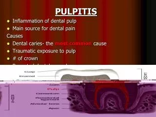
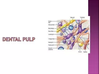
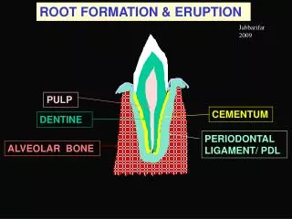
RESULTS and DISCUSSION • The extracellular matrix is the major constituent of the connective tissue. This is composed of ground substance and fibrillar proteins. The main cells of the connective tissue are the fibroblasts. The pulp also contains odontoblasts (the highest differentiated cells), undifferentiated mesenchyme cells, and immunocompetent cells (lymphocytes, macrophages, leucocytes).
Adequate pulp preparation has always been a challenge, because artefacts resulting from inadequate fixation often are described as evidence of pathosis. Methods with dropping a tooth in a jar of formalin, even if done immediately after extraction, are inadequate to permit subsequent critical examination of the dental pulp. Other methods are with section the apical 2–3 mm of the root with a fissure bur, and an opening was made into the pulp chamber with a round bur and fixed with 10% buffered formalin.
CONCLUSION • The developments of models are needed for better investigation of deciduous dental pulp, but also for better her preservation
Thank you for your attention ANY QUESTIUONS ?