Download
1 / 61
610 likes | 894 Views
Connective Tissues (generic) Digital Laboratory. It’s best to view this in Slide Show mode, especially for the quizzes. This module will take approximately 60 minutes to complete. After completing this exercise, you should be able to:

E N D
Connective Tissues (generic) Digital Laboratory It’s best to view this in Slide Show mode, especially for the quizzes. This module will take approximately 60 minutes to complete.
After completing this exercise, you should be able to: • Distinguish, at the light microscope level, each of the following components of connective tissue: • Collagen fibers • Elastic fibers (may need special stains) • Reticular fibers (seen with special stains only) • Fibroblasts • Mast cells (distinguished only with special stains - metachromasia) • Ground substance • Distinguish, at the light microscope level, each of the following types of connective tissue: • Loose (areolar) • Dense irregular • Dense regular (e.g. tendon) • Elastic tissue • Distinguish, at the electron microscope level, each of the following components of connective tissue: • Collagen fibers • Elastic fibers • Fibroblast • Basal lamina
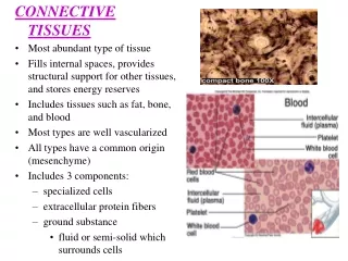
Before analyzing connective tissue (CT) in detail, it helps to first recognize that a tissue is indeed some form of CT. Unlike epithelia, which are composed of many cells with little extracellular matrix, CTs typically are composed of a few cells, with lots of extracellular matrix. On an H&E slide, this is readily apparent by comparing the number of nuclei in epithelia and CT: K = keratinized, but you already knew this epithelium connective tissue nuclei Border between epithelium and connective tissue
Two more examples of epithelia and connective tissue. It helps to remember that epithelia line a lumen, or space. Also note that epithelial cells tend to have more cytoplasm, which stains eosinophilic, while connective tissue cytoplasm is pale or sparse. a space epithelium connective tissue another space epithelium connective tissue
A little easy, but just in case, which areas of the slide are epithelial, and which are connective tissue? (advance PPT for answers) connective tissue epithelium Yeah we cropped away the space…OOOOOOH tricky.
Another one for the ages… the space Epithelium is between the dashed blue lines. Connective tissue is everything else (except the space). Note the area to the lower left with a number of nuclei. This is an area of connective tissue infiltrated with white blood cells.
Lets look at some of the components of connective tissue. • FIBERS: collagen fibers are thick and pink (purple block arrows) • reticular fibers, are present, but not visible without special stains • elastic fibers are thin and purple (black block arrows; not visible with standard H&E, but here stained with resorcin) • CELLS: A fibroblast is indicated; note its tapered appearance. • To the right, note the cells in military formation; these are probably white blood cells. • MATRIX: The clear spaces are filled with ECM, and in life contain water, ions, GAGs, etc., also called ground substance fibroblast cytoplasm and nucleus
Fibers of connective tissue - collagen COLLAGEN FIBERS Two more views of connective tissue, focusing on collagen fibers, which appear as pink threads. Note the thickness and abundance of collagen fibers varies. The purple arrows indicate collagen fibers cut in cross section. The top of the left image shows an undulating blood vessel.
Fibers of connective tissue - collagen In this electron micrograph from connective tissue, note the collagen fibers in longitudinal section (red circle) and in cross section (green circle).
Fibers of connective tissue – reticular fibers Although the proteins making up reticular fibers are homologous to those that make up collagen fibers, reticular fibers are much thinner, and, therefore, are not visible in routine H&E stained images (left). The numerous purple dots are nuclei of lymphocytes. Special stains, e.g. silver stains, are used to visualize reticular fibers (right, yellow arrows).
Fibers of connective tissue – elastic fibers 0 Elastic fibers are also fairly thin, and often do not show up well on routine H&E sections. However, in tissues such as large elastic arteries, they form prominent sheets that are readily apparent on H&E (left image, blue arrows) or with special stains (right image, resorcin stain, yellow arrows)
Fibers of connective tissue – elastic fibers microfibrils 0 elastin core In this electron micrograph from connective tissue, note the amorphous elastic tissue (green circle), consisting of dark microfibrils within a pale elastin core.
Fibers of connective tissue Video of connective tissue spread showing collagen and elastic fibers – SL1 • Link to SL 001 • Be able to identify: • Collagen fibers • Elastic fibers
Fibers of connective tissue Video of salivary gland showing collagen fibers – SL91 • Link to SL 091 • Be able to identify: • Collagen fibers • Simple cuboidal epithelium (review) • Simple columnar epithelium (review) • Stratified cuboidal/columnar epithelium (review)
Fibers of connective tissue Video of hilus (root) of lung showing collagen fibers – SL24 • Link to SL 024 • Be able to identify: • Collagen fibers • Pseudostratified epithelium (review, very poor prep) • Serous glands (review) • Mucous glands (review)
Fibers of connective tissue Video of elastic artery (H&E) showing elastic fibers – SL31 • Link to SL 031 • Be able to identify: • Elastic fibers
Fibers of connective tissue Video of elastic artery (H&E) showing elastic fibers (resorcin stain) – SL184 • Link to SL 184 • Be able to identify: • Elastic fibers
Fibers of connective tissue Video of spleen showing reticular fibers (silver stain) – SL32 Note: Resorcin staining of elastic fibers, and silver stains for reticular fibers, both show black threads that appear similar. While it is true that the elastic fibers shown here are thicker, that is not always the case. Therefore, for these special stains, you will be told what fibers are being highlighted. Note that this does not apply to the connective tissue spread (SL1), in which you should recognize collagen and elastic fibers. • Link to SL 032 • Be able to identify: • Reticular fibers
CONNECTIVE TISSUE ORIGIN Almost all connective tissues and some other tissues are derived from an embryonic precursor tissue called mesenchyme. This tissue is composed of mesenchymal cells surrounded by a matrix with very few fibers.
CONNECTIVE TISSUE ORIGIN Video of umbilical cord showing mesenchyme – SL35 Note: Mesenchyme is characterized by a near absence of connective tissue fibers, while loose connective tissue (shown later in this module) has some collagen and elastic fibers (though less than dense connective tissues). • Link to SL 035 • Be able to identify: • Mesenchyme • Mesenchymal cells
CELLS OF CONNECTIVE TISSUE - FIBROBLASTS Fibroblasts are derived from mesenchymal cells, and are the “resident” generic connective tissue cells that produce most, if not all, of the fibers and matrix. fibroblast cytoplasm and nucleus
CELLS OF CONNECTIVE TISSUE - FIBROBLASTS Video of connective tissue spread showing fibroblasts - SL1 • Link to SL 001 • Be able to identify: • fibroblasts
CELLS OF CONNECTIVE TISSUE - FIBROBLASTS Video of salivary gland showing fibroblasts – SL91 • Link to SL 091and SL 024 • Be able to identify: • fibroblasts
CELLS OF CONNECTIVE TISSUE - FIBROBLASTS 0 In electron micrographs, fibroblasts are spindle-shaped cells. Active fibroblasts will have a good amount of RER and Golgi necessary for synthesis of the extracellular fibers that the secrete. Quiescent fibroblasts will have less RER and Golgi.
CELLS OF CONNECTIVE TISSUE - FIBROBLASTS 0 Side-by-side images showing a light micrograph, and an EM corresponding approximately to the region within the box.
CELLS OF CONNECTIVE TISSUE - IMMUNE 0 Most of the other cells in connective tissues are not permanent residents, but immune cells that are “visiting”.
CELLS OF CONNECTIVE TISSUE - IMMUNE Most of the other cells in connective tissues are not permanent residents, but immune cells that are “visiting”. The image to the left is connective tissue; most of the cells you see are fibroblasts (green arrows). To right image is inflammation; this tissue used to look like the left image, but white blood cells, notably plasma cells (red arrows) and neutrophils (orange arrows) have infiltrated in response to an infection.
CELLS OF CONNECTIVE TISSUE - IMMUNE Video of inflammation – SL125 • Link to SL 125 • Be able to identify: • Nothing specific now, just a preview to inflammation, which you will learn about later
CELLS OF CONNECTIVE TISSUE - IMMUNE Macrophages are derived from monocytes and are found in many tissues throughout the body. The name (big eater) aptly describes their characteristic phagocytic capacity. Macrophages play roles in inflammation and in immune responses. Macrophages
CELLS OF CONNECTIVE TISSUE - IMMUNE Mast cells are immune cells that play a role in inflammation, and have secretory granules (arrows) containing histamine and heparin.
CELLS OF CONNECTIVE TISSUE - IMMUNE 0 In this specimen stained with the dye toluidine blue, many structures are stained blue. However, the components of the mast cell granules turn the dye a purple color (the color change of the dye is called metachromasia).
CELLS OF CONNECTIVE TISSUE - IMMUNE Video of connective tissue spread showing mast cells – SL118 • Link to SL 118 • Be able to identify: • Mast cells (FYI, the color on this digital slide is not optimal, but you can see mast cells if you look carefully.)
GROUND SUBSTANCE OF CONNECTIVE TISSUE The ground substance of connective tissue is the extracellular matrix (ECM) component that fills in the spaces between the fibers and cells. It contains water, ions, GAGs, and other molecules. The components vary considerably, and account for the fact that matrix (and connective tissues) can vary from fluid (left, also blood plasma) to solid (right).
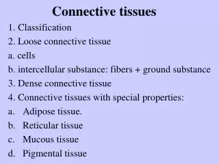
CLASSIFICATION OF CONNECTIVE TISSUE Connective tissue can be classified into generic and specialized : Generic connective tissues --Loose (aka areolar, includes reticular, elastic, maybe mesenchyme) --Dense irregular (aka dense irregular) --Dense regular (aka dense regular) We care about these today Specialized connective tissues --adipose --cartilage (hyaline, elastic, fibrous) --bone --blood These will be covered later.
It’s all about the extracellular stuff Generic connective tissues --Loose (aka areolar, includes reticular, elastic, maybe mesenchyme) --Dense irregular (aka dense irregular) --Dense regular (aka dense regular) If we overanalyze this a bit, the two variables we are using to differentiate these tissues are: 1. the density and thickness of the extracellular fibers (loose vs. dense) 2. the orientation of the fibers (irregular vs. regular) Of course, if we have two variables, with two possibilities each, that should leave us with a fourth generic connective tissue type, which would be “loose regular”. As we will see, from a functional standpoint, this tissue type would be ridiculous. Note that loose connective tissue is irregular, even though we don’t say this as part of the name because loose regular does not exist.
CLASSIFICATION OF CONNECTIVE TISSUE Loose connective tissue (aka areolar) appearance – more space than collagen or reticular fibers, fibers thin, so it is loose fibers oriented in all directions (but we don’t say irregular, remember?) function – not very strong, but usually is well vascularized. Found as filler, especially useful right under epithelia, which has no blood vessels of its own. (this is not H&E, but is a special stain similar to H&E that shows elastic fibers as black threads).
CLASSIFICATION OF CONNECTIVE TISSUE Fibroblast nuclei (fibroblast cytoplasm hard to see) the space collagen more Loose connective tissue (aka areolar) (everything that’s not epithelium) appearance – more space than collagen or reticular fibers, fibers thin, so it is loose fibers oriented in all directions (but we don’t say irregular, remember?) function – not very strong, but usually is well vascularized. Hey look, it’s right “under” the epithelium!!!!
CLASSIFICATION OF CONNECTIVE TISSUE Well, I’ll be darned if all these epithelia don’t have loose connective tissue right underneath them. Must be a good strategy for delivering nutrients to the epithelia, which were too stupid to have their own blood vessels. (Either that, or the blood vessels would disrupt the epithelial tissues‘ “barrier” quality.)
CLASSIFICATION OF CONNECTIVE TISSUE All connective tissues vary to some extent in the amount of collagen fibers, reticular fibers, and elastic fibers. This is especially true for the loose connective tissues. These variations give these tissues slightly different functional features, and in some cases, alternate names. Reticular tissue – the reticular fibers in this special stain are black. (nuclei are red) Elastic tissue – the elastic fibers are the bright eosinophilic bands you see. Though the elastic fibers look a little like collagen from the previous slides, elastic fibers tend to be “shinier”, especially when you lower the condenser on your microscope. Both of these are really cute variations of loose connective tissue
CLASSIFICATION OF CONNECTIVE TISSUE Mesenchyme is undifferentiated connective tissue that can give rise to all connective tissue types, including the generic types as well as the specialized ones (e.g. cartilage, bone, etc.). Because it has few extracellular fibers, it’s tempting to call this loose connective tissue. DON’T DO THIS!!!. And don’t call loose connective tissue mesenchyme. They don’t get along, and are very insulted when you do this. Although mesenchyme is EXTREMELY cool when discussing Embryology, and we do look at it in Histology, it probably won’t appear on your exam because it’s too tricky.
CLASSIFICATION OF CONNECTIVE TISSUE OK, back to our classification scheme. More pictures… Fibroblast nuclei Collagen fibers: Longitudinal Cross section oblique Dense irregular connective tissue appearance – less space, many (type I) collagen fibers, many are thick, so it is dense collagen fibers oriented in all directions, so it is irregular function – strong in multiple directions, but usually is not as well vascularized as loose
CLASSIFICATION OF CONNECTIVE TISSUE Video of salivary gland showing loose and dense irregular connective tissues – SL91 • Link to SL 091 • Be able to identify: • Loose connective tissue • Dense irregular connective tissue
CLASSIFICATION OF CONNECTIVE TISSUE Video of salivary gland showing loose and dense irregular connective tissues – SL125 • Link to SL 125 • Be able to identify: • Loose connective tissue • Dense irregular connective tissue
CLASSIFICATION OF CONNECTIVE TISSUE Fibroblast nuclei Collagen fibers (basically everywhere) Dense regular connective tissue appearance – no space, many thick (type I) collagen fibers packed so tightly, it’s hard to see individual fibers, so it is dense collagen fibers oriented in one direction, this is difficult to appreciate because the individual collagen fibers are hard to see, but look how the fibroblast nuclei have to orient and narrow to accommodate, so it is regular function – strong in one direction (e.g. tendon, ligament), but usually very poorly vascularized, so it takes a long time to heal
CLASSIFICATION OF CONNECTIVE TISSUE Video of tendon showing dense regular connective tissue – SL27 • Link to SL 027 • Be able to identify: • Dense regular connective tissue
The next set of slides is a quiz for this module. You should review the structures covered in this module, and try to visualize each of these in light and electron micrographs. • Distinguish, at the light microscope level, each of the following components of connective tissue: • Collagen fibers • Elastic fibers (may need special stains) • Reticular fibers (seen with special stains only) • Fibroblasts • Mast cells (seen in special stain stains only - metachromasia) • Ground substance • Distinguish, at the light microscope level, each of the following types of connective tissue: • Loose (areolar) • Dense irregular • Dense regular (e.g. tendon) • Elastic tissue • Distinguish, at the electron microscope level, each of the following components of connective tissue: • Collagen fibers • Elastic fibers • Fibroblast • Basal lamina
Final quiz Self-check: Identify the tissue. (advance slide for answers) Dense irregular connective tissue
Final quiz Self-check: Identify the tissue. (advance slide for answers) dense regular connective tissue
Final quiz Self-check: Identify cells A and B. Identify structure at arrows and . (advance slide for answers) Epithelial cells A A Basal lamina fibroblasts B Reticular (collagen) fibers B
Final quiz Self-check: This tissue had been previously injured. What is the significance of the structure at the arrow? (advance slide for answer) Synthesis of a new basal lamina.