Download
1 / 11
110 likes | 214 Views
New Technology for Protein Separation. BME 273 Cathy Castellon Advisor: Dr. Rick Haselton Graduate Advisor: Greg Stone. Protein Background.

E N D
New Technology for Protein Separation BME 273 Cathy Castellon Advisor: Dr. Rick Haselton Graduate Advisor: Greg Stone
Protein Background • To compare the expression of protein profiles from an arbitrary reference state of a cell, tissue, or organism, to the profile of an non-standard condition • Master Molecules of Living Things • Composition • Central Dogma
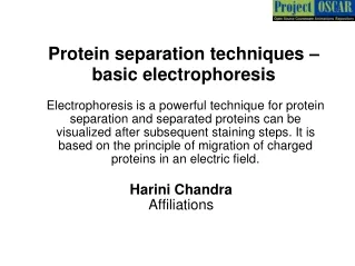
How Does Current Technology Work? • Separates proteins by: • isoelectric point (pI) • size (molecular weight) • Problems • large volume • time consuming • resolution problems • labor intensive • Economics • Need a faster more efficient technology that will separate proteins 100-fold at once
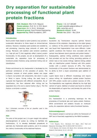
Design Goals • Adapt Micro-fluid Technique • Create flow channel (soft lithography) • Separation based on hydrophobicity • Create inlet and outlet points • Load fluorescently labeled protein solution into one end • Pump buffer solution through the channel • Fluoremeter will detect separation • Automate
Lithography Technique • Coat Substrate with Photoresist • Apply Mask/ Expose Photoresist to Light • Develop Photoresist • Cast and Cure PDMS • Remove PDMS from Substrate
Detailed Channel Design • 2X2 cm lanes • Hydro-phobic/phyllic on same slide (R,L) • Posts used to aid mixing and accentuate the separation
Slides 3-glicidoxypropyltrimethoxysilane octyltrichlorosilane • Gradient • hydro-phobic/phyllic • Contact Angle Measurements • PDMS Adherence
Strategy for Prototype Lysozyme Cytochrome C • 2 different proteins • CytochromeC and Lysozyme • 2 different labels • AlexaFluro 430 (540nm) and 350 (442nm)
Unforeseen Problems • Si-Lanes lost reactivity • Gradient could not be improved • Micro-fluid channel • leak • HPLC column • Incorrect size
Future Work • Flow Chamber-basic idea • monitor pressure of flow • monitor flow rate • Spectrophotometer • test each reservoir and measure labeled protein signal
References • DoInik, Vladislav, Shaorong Liu, and Stevan Jovanovich. Capillary electrophoreses on microchip. Electrophoresis 2000. 21, 41-54. • Stroock, Abraham D., Stephan K.W. Dertinger, etal. Chaotic Mixer for Microchannels. Science. Vol 295, 647-651. • Hopp, Thomas P. and Kenneth R. Woods. Prediction of Protein Antigenic Determinants from Amino Acid Sewquences. National Academy of Sciences of the USA. Vol. 78, Issue 6, 3824-3828. • http://www.sdk.co.jp/shodex/english/dc010603.htm • http://mstflab.vuse.vanderbilt.edu/projects/microfluidics/soft_lithography_intro.html • http://www.unitedchem.com/1024x768/Uct2.htm • http://metallo.scripps.edu/PROMISE/1BBH.html • http://www.rcsb.org/pdb/molecules/pdb9_1.html • http://www.worthington-biochem.com/manual/L/LY.html • http://crystal.uah.edu/~carter/protein/xray.htm • Acknowledgements • Dr. Rick Haselton, Advisor, Vanderbilt University • Greg Stone, Graduate Student, Vanderbilt University • David Schaffer, Graduate Student, Vanderbilt University • Dr. David Hachey, Mass Spectrometry Vanderbilt University