Download

1 / 21
220 likes | 361 Views
FREE LIVING AMEBAE. LECTURER: SR. NORAZSIDA. INTRODUCTION. A large and diverse group of protozoan organisms. Inhabit fresh and salt water. Decaying organic matter and damp soil. 2 potential pathogens r: 1) Naegleria fowleri 2) Acanthamoeba sp. N. fowleri. Morphology.

E N D
FREE LIVING AMEBAE LECTURER: SR. NORAZSIDA
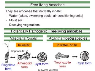
INTRODUCTION • A large and diverse group of protozoan organisms. • Inhabit fresh and salt water. • Decaying organic matter and damp soil. • 2 potential pathogens r: 1) Naegleria fowleri 2) Acanthamoeba sp.
Morphology • Family Vahlkampfiidae • Ameboflagellates – having both an ameboid and flagellate stage in their life. • Agent of primary amebic meningoencephalitis (PAM) or Naegleriasis. • Since 1965, 140 cases have been reported world wide. • Risk group: healthy children and young adults with a history of swimming or diving in fresh or brackish water. • Sources of infection: contaminated swimming pools, stagnant ponds, freshwater lakes and streams, thermal springs and spas.
Drought and elevated temperatures increase concentrations of N. fowleri. Why? Bcoz, they feed on large populations of bacteria in these warmed water souces. • Transmitted via inhalation of contaminated dust. • Transmission: nasal instillation follows olfactory nerve to CNS • Trophozoite : 7-20µm, large, broad pseudopods, single nucleus, large central karyosome.
Life cycle • Consists of 3 stages: 1) an amebic trophozoite (the only stage that exist in human) 2) a biflagellate form 3) cyst
PATHOGENESIS • The incubation period is generally 3-7 days accompanied by the prodromal symptoms of headache and fever. • Rapidly progress to frank meningitis with the onset of nausea and vomiting (stiff neck), confusion and coma. • Death usually occurs in 3-6 days following the onset of these serious symptoms.
Primary amebic meningoencephalitis PAM • 1-14 days incubation period • symptoms usually within a few days after swimming in warm still waters • infection believed to be introduced through nasal cavity and olfactory neuroepithelium • symptoms include headache, lethargy, disorientation, coma • rapid clinical course, death in 4-5 days after onset of symptoms • trophozoites can be detected in spinal fluid, but diagnosis is usually at autopsy
LABORATORY DIAGNOSIS • Finding motile trophozoites in: -saline and wet preps of fresh spinal fluid -nasal discharge -Tissue biopsy • Induced within 2-20 hours by transferring the ameboid form from tissue or CSF to water and incubating at 370C. • Cultivation technique • PCR • Indirect fluorescent antibody procedures.
TREATMENT • Amphotericin B • Combination treatments: • Amphotericin B + miconazole • Amphotericin B + rifambin
PREVENTION • Public and private swimming pools, hot tubs, and baths should be properly maintained and adequately chlorinated to prevent growth of the organism.
Morphology • Family of Ancanthamoebidae. • Never produce flagella. • Produce a chronic infection of the CNS: Granulomatous amebic encephalitis (GAE). • Also caused a keratitis and skin ulcers. • Risk group: immunocompromised, chronically ill. • Transmission: respiratory or skin with hematogenous spread to CNS • Trophozoite: 10-45µm, spiny acanthopodia, single nucleus, large central karyosome • Cyst: 10-20µm, rounded, double walled (outer wall having a wrinkled appearance), single nucleus.
Life cycle • Consists of: • 1) trophozoite 2) cyst. • resistant to dessication and mild chlorination. • Carried out by water and through the air.
PATHOGENESIS • GAE has an insidious onset. • CNS infection is acquired hematogenously by the inhalation/aspiration of trophozoites and cysts caused pneumonitis. • Or through skin and mucosal ulceration. • Incubation period: questionable, may take weeks to months to progress. • Slow process of tissue invasion tends to stimulate granuloma formation. • Single or multiple focal lesions develop over a prolonged period marking the chronic nature of the disease. • Symptoms gradually develop: headache, fever, fatigue, stiff neck, and altered mental status.
Granulomatous Amebic EncephalitisGAE • portal of entry unknown, possibly respiratory tract, eyes, skin • presumed hematogenous dissemination to the CNS • infection associated with debility or immunosuppression • onset is insidious with headache, personality changes, slight fever • progresses to coma and death in weeks to months • amebas not yet detected in spinal fluid
LABORATORY DIAGNOSIS • By finding motile trophozoite in spinal fluid specimen. • By finding trophozoite and cyst form in: -brain biopsy tissue histological examination -scrapings from cutaneous or corneal lesions. • Indirect immunofluorescent staining techniques. • Cultured on non-nutrient agar, overlaid with viable E.coli bacteria stained with Giemsa or calcofluor white identification of cyst or trophozoite.
TREATMENT & PREVENTION • Sulfamethazine; however, most cases are only diagnosed at autopsy. • no human cures documented • Symptoms develop gradually over a prolonged period of time the disease may be overlooked.
AMEBIC KERATITIS • predisposing factors • ocular trauma • contact lens (contaminated cleaning solutions) • symptoms • ocular pain • corneal lesions (refractory to usual treatments) • diagnosis • demonstration of amebas in corneal scrapings • treatment • difficult, limited success • corneal grafts often required