Download
1 / 41
470 likes | 1.1k Views
Acute Kidney Injury. Charles Pizanis , MD Division of Hospital Medicine Department of Internal Medicine 7 August 2014. Objectives. Become aware of the definitions, general epidemiology, and prognosis of acute kidney injury

E N D
Acute Kidney Injury Charles Pizanis, MD Division of Hospital Medicine Department of Internal Medicine 7 August 2014
Objectives • Become aware of the definitions, general epidemiology, and prognosis of acute kidney injury • Identify the indications, utility, and limitations of commonly used diagnostics • Review several select causes of acute kidney injury • Go over several practical pointers
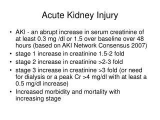
Definitions • RIFLE Criteria • Risk – 1.5-fold increase in creatinine over not more than 7 days OR GFR decrease by 25% OR urine output <0.5ml/kg/hr for 6 hours • Injury – 2-fold increase in creatinine over not more than 7 days OR GFR decrease by 50% OR urine output <0.5ml/kg/hrfor 12 hours • Failure – 3-fold increase in creatinineover not more than 7 days OR GFR decrease by 75% OR urine output <0.3ml/kg/hr for 24 hours OR anuria for 12 hours • Loss – complete loss of kidney function for more than 4 weeks • ESRD - complete loss of kidney function for more than 3 months • AKIN Criteria • Acute kidney injury – within 48 hours - absolute increase in creatinine ≥ 0.3 mg/dLOR increase in creatinine by ≥ 50% OR urine output of <0.5ml/kg/hr for > 6 hours • Staging Criteria (AKIN Stage 1, 2, 3) • KDIGO Criteria • Acute kidney injury – absolute increase in creatinine ≥ 0.3 mg/dL within 48 hours OR ≥ 1.5-fold increase in creatininebaseline known or presumed to have occurred within seven days OR urine output of <0.5ml/kg/hrfor > 6 hours • Staging Criteria (KDIGO Stage 1, 2, 3)
Differential • Prerenal • Hypovolemia • Hypotension • Cardiorenal syndrome • Hepatorenal syndrome • Renal artery stenosis • Medications (ACE inhibitors, NSAIDs, calcineurin inhibitors) • Intrinsic Renal • Vascular • Hypertensive emergency • Thrombotic thrombocytopenic purpura-hemolytic uremic purpura • Thromboembolic disease • Glomerular • Glomerulonephritides • Glomerulonephropathies • Tubular/Interstitial • Acute tubular necrosis • Acute interstitial nephritis • Tumor lysis syndrome • Pigment induced nephropathy • Post-Renal • Benign prostatic hypertrophy • Prostate cancer • Bladder cancer • Stones • Bladder dysfunction • Retroperitoneal fibrosis
How Common is AKI? • 2001 National Hospital Discharge Survey • 29,039,599 hospitalizations • 558,032 cases of AKI • 19.2 per 1000 hospitalizations • LOS 2 days longer • OR 2.0 discharge to SNF or inpatient rehab • OR 4.1 in-hospital mortality
Hospital Setting • Madrid multicenter tertiary care study • ATN – 45% • Prerenal – 21% • Acute on chronic kidney disease – 13% • Obstructive – 10% • Glomerulonephritis – 3% • AIN – 2% • Vasculitis – 2% • Vascular – 1% • Karachi tertiary care study • Prerenal – 70% • Intrinsic renal – 22% • Post-renal – 8%
Prognosis Cerda 2008
Prognosis – AKI Leading to CKD Coca 2012
Prognosis – AKI Leading to ESRD Coca 2012
Prognosis – AKI Leading to Death Coca 2012
Prognosis – Sequential AKI Leading to Death Thakar 2011
Objectives • Become aware of the definitions and general epidemiology and prognosis of acute kidney injury • Identify the indications, utility and limitations of commonly used diagnostics • Review several select causes of acute kidney injury • Go over several practical pointers
Diagnostics • History and physical exam • Basic metabolic panel • Urinalysis • Urinalysis with reflex to culture • Urine microscopy • Fractional excretion of sodium, urea • Urine eosinophils • Fluid challenge • SPEP, UPEP, Light chains • Uric acid • Bladder scan (post-void residual) • Retroperitoneal ultrasound • Bladder pressure • Computed tomography (CT) • Transthoracic echocardiogram
History and Physical Exam Items of Use • History • Volume losses – diarrhea, vomiting, decreased PO, bleeding • Urinary symptoms – dysuria, urgency, decreased output, intermittent stream, flank pain • Medications – new or changes in dosing • Recent procedures, imaging • Vitals • Fever • Heart rate • Blood pressure (including orthostatics) • Physical Exam • Volume assessment • Stigmata of liver disease • Genitourinary exam
Basic Metabolic Panel • Electrolyte, acid base disturbance • BUN/Creatinine • >20:1 indicative of prerenal • Sensitivity 81%, Specificity 92% • Caveats • Elevations in BUN caused by GI blood loss, corticosteroid use • Low creatinine in low muscle mass
Urinalysis • Specific gravity • pH • Blood • Protein • WBCs, Leukocyte esterase • RBCs • Nitrites • Urinalysis reflex to culture if suspect infection
Urine Microscopy • Use: identifying casts, dysmorphic RBCs, urinary sediments • Granular “muddy brown” casts indicates ATN • Sensitivity 82%, Specificity 72% • Acanthocytes (dysmorphicrbcs) indicates glomerulonephritis • Sensitivity 62%, Specificity 89% • WBC casts indicates kidney inflammation (pyelo, AIN) • Caveats • Nephrology consultation generally recommended
http://renalfellow.blogspot.com/2009/06/acanthocyte.html http://www.agora.crosemont.qc.ca/urinesediments/Imdoceng/d21d002.html
Fractional Excretion of Sodium (FeNa) • Use: differentiating prerenal from ATN • FeNa < 1% indicates prerenal • Sensitivity 77%, Specificity 96% • FeNa >2 indicates ATN • Sensitivity 72%, Specificity 88% • Caveats • Diuretic use – associated with increased FeNa • Sensitivity 48% • Alkalosis – associated with increased FeNa • Myoglobinuria, contrast nephropathy, acute glomerulonephritis, post-renal causes – associated with decreased FeNa • FeNa between 1 and 2?
Fractional Excretion of Urea (FeUrea) • Use: identifying prerenal from ATN • FeUrea < 35% indicates prerenal • Sensitivity 85%, Specificity 92% • FeUrea > 50% • Sensitivity 32%, Specificity 82% • Caveats • Mannitol, acetazolamide – associated with increased FeUrea • FeUrea between 35 and 50?
Urine Eosinophils • Use: identifying acute interstitial nephritis, cholesterol emboli • Sensitivity 30-63%, specificity 68-91% • Caveats • Seen also in pyelonephritis, prostatitis, RPGN
Fluid Challenge • Use: identifying prerenal cause • Sensitivity, specificity not well documented • Caveats • Correlation does not prove causation! • How much fluid is adequate?
Bladder Scan (Post-void Residual) • Use: identifying urinary retention • Bladder scan or in-and-out catheterization • Caveats • No absolute value • Ascites, cystic ovarian disease, pregnancy falsely elevate bladder scan http://www.medicalexpo.com/prod/verathon-medical-europe/portable-ultrasound-bladder-scanners-70452-530575.html
Retroperitoneal Ultrasound • Use: identifying anatomic abnormalities of GU system • Non invasive testing • Caveats • Obese patients
Bladder Pressure • Use: identifying elevated intraabominal pressures (abdominal compartment syndrome) • Sustained bladder pressure ≥ 12 mmHg defines intraabominal hypertension • Sustained bladder pressure > 15 mmHg indicates compartment syndrome • Sensitivity 56%, Specificity 87% • Caveats • Patient must have foley • High variability in technical skill http://www.uptodate.com/contents/abdominal-compartment-syndrome?source=search_result&search=bladder+pressure&selectedTitle=1%7E1
Objectives • Become aware of the definitions and general epidemiology and prognosis of acute kidney injury • Identify the indications, utility and limitations of commonly used diagnostics • Review several select causes of acute kidney injury • Go over several practical pointers
Mr. R • 52-year-old male comes in at request of PCP after “abnormal lab results.” Creatinine is 2.8 mg/dL with baseline of 0.8 mg/dL. Patient endorses fatigue but other ROS is negative. Patient’s only past medical history is alcoholic cirrhosis for which he is on no medications. • Evaluation including urinalysis, urine microscopy, bladder pressures, renal ultrasound, fluid challenge are all non-revealing. • What is a likely diagnosis?
Hepatorenal Syndrome (HRS) • Syndrome of acute kidney injury in setting of portal hypertension • Pathophysiology: arterial vasodilitation in splanchnic vessels -> decreased SVR -> renal arterial vasoconstriction -> decreased GFR, increased Cr • Type 1 HRS: doubling of creatinine to >2.5 mg/dL in less than two weeks • Type 2 HRS: less severe than type 1 • Diagnosis of exclusion • “Albumin challenge” – 1 g/kg daily for at least two days and hold diuretics • Treatments • ICU – norepinephrine + albumin • Non-ICU – midodrine (7.5 mg PO TID) + octerotide (100 mcg subq TID x 2 doses then 200 mcg subq TID) + albumin • Consider TIPS if failing • Palliative Care
Ms. S • 62-year-old female with history of HIV-negBurkitt lymphoma s/p cycle 1 of induction chemo six days prior. Patient complains of fatigue, dyspnea but otherwise negative ROS. Initial evaluation reveals the following labs: Ca 6.8 98 48 138 Mg 1.8 6.2 2.9 (baseline 1.0) 28 Phos7.8 What is a likely diagnosis?
Tumor Lysis Syndrome (TLS) • Syndrome of hyperkalemia, hyperphosphatemia, hyperuricemia, hypocalcemia from cell lysis in setting of malignancy and/or treatment of malignancy often with resultant end organ damage • Risk factors: large tumor burden, certain tumor types (hematologic malignancy), recent cytotoxic therapy • Pathophysiology • Xanthinuria, hyperuricema: excess cell turnover -> purine catabolism -> hyperxanthinuria, hyperuricemia-> xanthine, uric acid crystalization in tubules • Hyperphosphatemia: excess cell turnover -> hyperphosphatemia -> calcium phosphate crystalization in tubules • Diagnosis • Laboratory TLS = 2 or more three days before or within seven days of therapy: hyperkalemia, hyperphosphatemia, hyperuricemia, hypocalcemia • Clinical TLS = laboratory TLS plus increased creatinine (≥1.5 upper limit of normal), cardiac arrythmia/sudden death, seizure not attributable to therapy • Treatment/Management • Hydrate, hydrate, hydrate • Diuretics • Hyperuricema – allopurinol OR rasbirucase – depending on uric acid levels • Trend BMP, Ca, Phos, uric acid, vitals, urine output • Hemodialysis – oliguria, anuria, persistent electrolyte disturbance
Trauma Alert Doe • 111-year-old male without known medical history admitted to MICU after being found down in own vomit. Hypotensive on presentation, unknown down time. Intubated for airway protection and on pressors. Chemistry as follows: UA: Spec grav1.08 pH 6.0 Blood large LCE moderate WBC 3 RBC 0 98 68 128 4.8 3.8 (baseline unknown) 16 What is a likely diagnosis?
Pigment Induced Nephropathy • Acute kidney injury in setting of excess heme-pigment molecules (myoglobin, hemoglobin) • Risk factors: rhabdomyolysis, hemolysis, volume depletion • CK levels > 5000 u/L OR = 5.12 • Pathophysiology: cell breakdown -> release of heme-pigment molecules -> tubular obstruction, direct tubular injury • Diagnosis: AKI in appropriate clinical setting • Suggestive UA, elevated CK, hemolytic labs • Treatment/Management • Hydrate, hydrate, hydrate – usually titrate to urine output of 200-300 mL/hr • Trend CKs, H/H, BMP, vitals, urine output
Objectives • Become aware of the definitions and general epidemiology and prognosis of acute kidney injury • Identify the indications, utility and limitations of commonly used diagnostics • Review several select causes of acute kidney injury • Go over several practical pointers
Practical Pointers - Medications • Creatinine clearance • Measured by Cockcroft-Gaultformula • Cockcroft-GaultCrCl = (140-age) * (Wt in kg) * (0.85 if female) / (72 * Cr) • USE IDEAL BODY WEIGHT!!! • DVT Prophylaxis • CrCl 30-50 mL/min • Fondaprinux 50% dose decrease or just use heparin • CrCl < 30 mL/min • Enoxaparin 30 mg daily or just use heparin • Fondaparinux contraindicated • ESRD – use heparin
Practical Pointers - Analgesia • NSAIDs, COX 1/2 inhibitors- avoid at all costs! • Aspirin has lowest effect on GFR • Acetaminophen generally safe • Total daily dose not to exceed 3 grams • Opioids – be cautious with them! • Most opioids undergo renal excretion primarily (methadone main exception) • Toxic effects usually related to metabolites • Dose reduction almost always indicated – 75% • Severe pain – fentanyl • Metabolites are non-toxic • Non-opioid adjuncts • Gabapentin – renally adjusted
Practical Pointers - Antibiotics • Antibiotics • Many are renally adjusted • Vancomycin IV • Initial bolus dose is weight based • Subsequent dose is based on renal function • ESRD on HD usually dosed after dialysis • Piperacillin/tazobactam IV • CrCl < 20 mL/min or ESRD – dosed every 12 hours
Practical Pointers – Contrast Studies • Gadolinium relatively contraindicated if CrCl <30-60 • Nephrogenic systemic fibrosis is bad • MRI without contrast • Iodine based contrast • Risk dependent on type and amount of contrast, GFR, comorbidities • CT studies • Prevention – IVF, avoidance of nephrotoxins • N-acetylcystine data controversial http://archderm.jamanetwork.com/article.aspx?articleid=712220
Practical Pointers – Renal Transplant Patients • Renal transplant patient admitted = automatic Transplant Nephrology consult • Helpful workup to get things started • Transplant kidney ultrasound – “US Renal Transplant w/ w/o doppler” • Calcineurin inhibitor serum trough levels • 12 hours after dose • Tacrolimus – “FK506-Tacrolimus-Lab” • Cyclosporine – “Cyclosporin-Lab” • Blood, urine BK virus PCR • If concerned for acute rejection, call Transplant ASAP http://www.arizonatransplant.com/healthtopics/
Practical Pointers – Use of PowerChart • Documentation • “Elevated creatinine” - “Renal failure, unspecified” • “Acute kidney injury” – “Acute renal failure” – “Acute on chronic renal failure” • Comment on cause – “with lesion of tubular necrosis” • “Chronic kidney disease” – 3 months • Comment on stage • Comment on cause • “End-stage renal disease” – 3 months • Order set for Acute Kidney Injury – “Adult Acute Kidney Injury” • Order set for Hepatorenalsyndrome – “Adult Hepatorenal Syndrome” • Favorites
Resources • Cerda J, et al. Epidemiology of acute kidney injury. Clin J Am SocNephro2008;3(3):881-886. • Coca SG, Singanamala S, Parikh CR. Chronic kidney disease after acute kidney injury: a systematic review and meta-analysis. Kidney Int2012;81:442-448. • Thakar CV, Christianson A, Himmelfarb J, Leondard AC. Acute kidney injury episodes and chronic kidney disease risk in diabetes mellitus. Clin J Am SocNephro 2011;9(7):2567-2572. • Liano F, Pascual J, and the Madrid Renal Failure Study Group. Epidemiology of acute renal failure: a prospective multicenter, community-based study. Kidney Int 1996;50:811-818. • Macedo E, Mehta RL. Prerenal failure: from old concepts to new paradigms. CurrOpinCrit Care 2009;15(6):467-473. • Carvounis CP, Nisar S, Guro-Razuman S. Significance of the fractional excretion of urea in the differential diagnosis of acute renal failure. Kidney Int 2002;62:2223-2229. • PerazellaMA, Coca SG, Kanbay M, Brewster UC, Parikh CR. Diagnostic value of urine microscopy for differential diagnosis of acute kidney injury in hospitalized patients. Clin J Am SocNephro2008;3(6):1615-1619. • Muriithi AK, et al. Utility of urine eosinphils in the diagnosis of acute interstitial nephritis. Clin J Am SocNephro 2013;8(11):1857-1862. • El-Abdellati E, et al. An observational study on rhabdomyolysis in the intensive care unit. Exploring its risk factors and main complication: acute kidney injury. Ann Intens Care 2013;3(8):1-8. • Pham PCT, et al. Pain management in patients with chronic kidney disease. Clin Kidney J 2009;2(2):111-118.