Download
1 / 68
760 likes | 1.13k Views
HEAD AND NECK CASE CONFERENCE (PAPILARY THYROID CARCINOMA). Philippine Academy for Head and Neck Surgery, Inc. – Medical Center Manila June 10, 2013 12 PM. General Data. R.M. 51 y/o Female. Chief Complaint. Anterior neck mass. History November, 2011.

E N D
HEAD AND NECK CASE CONFERENCE (PAPILARY THYROID CARCINOMA) Philippine Academy for Head and Neck Surgery, Inc. – Medical Center Manila June 10, 2013 12 PM
General Data • R.M. • 51 y/o • Female
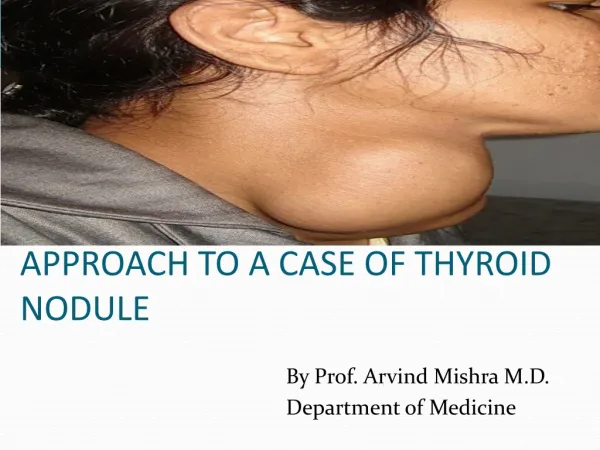
Chief Complaint • Anterior neck mass
History November, 2011 • Notable mass at the left anterior neck approximately 2x3x3 cm • Physical Examination: • (+) 3X2X2 cm cervical lymphadenopathy, post-auricular area • (+) 2x3x2 cm left anterior neck mass, firm, moves with deglutition
What would be your diagnostic work-ups ? • How do you do it in your institution or practice?
Ultrasound of the neck10/26/11 • The right thyroid lobe is normal in size and measures 4.98 x 1.69 x 1.89 cm. While the left thyroid lobe is slightly enlarged and measures 5.60 x 2.14 x 2.20cm • There are lobulatedhypoechoic lesions in both thyroid lobes with location and sizes: • Right: • Mid portion (2 nodules) = 0.36 x 0.35 x 0.33 cm and 1.42 x 0.76 x 0.93 cm • Left • Upper to mid portion with numerous punctate calcification in the margins • A solitary enlarged hypoechoic lymph node with thickened cortex and intact fatty hilum is noted in the left lateral neck measuring 1.87 x 1.73 x 1.0 cm
Ultrasound of the neck10/26/11 • Impression; • Normal sized right thyroid lobe and enlarged left lobe with solid nodules. The lesion in the left lobe is vascular with calcifications. • Reactive left cervical adenopathy.
FNAB cytology Report11/2/2011 • Organ for aspiration biopsy: left thyroid and left cervical lateral node • Cytologic Diagnosis: • Cell findings are consistent with a papillary carcinoma of the thyroid, left, with metastasis to the left lateral neck area. • Cytologic Description: • Aspirate smears from all slides (4) appear similar and show clusters of atypical thyrocytes forming papillary patterns, there is modest colloid in the background. • There are also histiocytes present. • The nuclei shows grooves and inclusions.
How do you interpret fnac /fnab results/ • Methesda scoring /nomenclature for fnab?
Impression? Stage? • Plan of Management?
THYROID CARCINOMA STAGING • all anaplastic carcinomas are considered T4 tumor American Joint Committee on Cancer (AJCC) Staging Manual, 7th edition (2010)
THYROID CARCINOMA STAGING American Joint Committee on Cancer (AJCC) Staging Manual, 7th edition (2010)
THYROID CARCINOMA STAGING American Joint Committee on Cancer (AJCC) Staging Manual, 7th edition (2010)
THYROID CARCINOMA STAGING American Joint Committee on Cancer (AJCC) Staging Manual, 7th edition (2010)
OperationNovember 8, 2011 • Total thyroidectomy with modified radical neck dissection Type III, left
Is there still a controversy between total and subtotal thyroidectomy? In this case? • Role of central neck dissection? • Types of neck dissection? Comprehensive or selective?
Record of OperationNovember 8, 2011 • Findings: • Thyroid gland enlarged • Right lobe 4x3x2 cm with solitary nodule 1 cm in diameter • Left lobe 5x3.5x3 with 2 nodules • #1 located at superior pole – 3x3x3cm • #2 located at inferior pole -1x1x1 cm • (+) enlarged cervical nodes/ jugular chain of nodes, left ~5 in number: 2 were dark-colored -2x2x2 cm in greatest dimensions, other 3 were light-colored
Record of OperationNovember 8, 2011 • Post-op Diagnosis: S/P Total Thyroidectomy, Modified Radical Neck Dissection Type III for Papillary Thyroid Carcinoma Stage IVA (sT2N1bM0)
Final HistopathNovember 8, 2011 • Papillary carcinoma, left and right lobes of the thyroid (2.3 cm, left lobe, and two foci in the right lobe, 0.2 cm and 0.4 cm) • Background of focal lymphocytic thyroiditis • Surgical lines of thyroidectomy are negative for tumor. • Positive for tumor metastasis to 9/18 left cervical LN
Whole body I-131 ScintigraphyDec. 17, 2011 • S/P RAI therapy (12/13/11) • Whole body scans were obtained 4 days after administration of a 100 mci oral therapeutic dose of I-131. • There are foci of dense tracer activity in the right and left thyroid beds representing uptake of the therapy dose by functioning residual thyroid tissues. These measured 1.2x1.2 cms and 1.6x1.6 cms, respectively. • Faint, ill-defined tracer localization is seen in the inferior thyroid bed likewise denoting residual functioning thyroid. • Physiologic tracer accumulation noted in the nasopharynx, salivary glands, gastrointestinal tract and urinary bladder. • No functioning metastasis appreciated.
Whole body I-131 ScintigraphyDec. 17, 2011 • Interpretation: Functioning thyroid tissue remnants limited to the anterior area.
What are the controversies in thyroid scanning ? • How do you give RAI? What are the doses?
2/13/12 • Levothyroxine 100mg OD
3/14/12 • Levothyroxine 100mg OD
4/13/12 • Levothyroxine 100mg OD
Role of TSH suppression? • How do you follow up? • Serum TSH • Serum thyroglobulin • Neck ultrasound • Prognosis?
7 months post-operative • No palpable neck mass • Persistently low TSH and elevated TG • Ultrasound of the neck was requested
Ultrasound 6/26/12 • Scan over the post-cervical bed shows subcentimeter hypoechoic nodular foci in the lower anterior and left para-tracheal region measuring 0.34 to -0.85 cm. lateral to the said nodule is a 1.19 cm lymph node at level V. • Subcentimeter lymph nodes with fatty hylum are demonstrated in both submandibular, submental and right jugular chain with sizes ranging from 0.19-0.74 cm. • The submandibular and parotid glands are intact. • Impression • S/P Total thyroidectomy from known papillary thyroid carcinoma with subcentimeter nodules in the lower anterior and left lateral neck and a slightly enlarged left cervical lymph nodes likely tumor recurrence. Unremarkable submandibular and parotid glands.
6/27/12 • Levothyroxine 100mg OD
Whole body I-131 scintigraphyOct 8, 2012 • S/P RAI therapy (10/03/12) • Whole body scans were obtained 5 days after administration of 150 mCi oral therapeutic dose of I 131 • There are confluent foci of ill-defined tracer activity in the thyroid beds representing uptake of the therapy dose by functioning residual thyroid tissues aggregate measurements were approximately 3 x 6 cms. • Physiologic tracer accumulation noted on the nasopharynx, salivary glands, GIT, and urinary bladder • No functioning metastasis is seen
Whole body I-131 scintigraphyOct 8, 2012 • Interpretation: Functioning thyroid tissue remnants limited to the thyroid bed.
12/10/12 • Levothyroxine 150g (mon- sat, 1/2 on Sunday)
Record of Operation 4/26/13 • Pre-operative diagnosis: Recurrent papillary thyroid cancer; S/p Total thyroidectomy with modified radical neck dissection Type III, left • S/P RAI 12/13/11 and 10/03/12
Record of Operation 4/26/13 • Operation: Central node dissection
Record of Operation 4/26/13 • Findings: multiple adhesions between strap muscle, trachea and surrounding areas, #1 enlarged LN ~ 1cm at Left paratracheal area, multiple persistent LN on central area 0.3-1cm in diameter adherent to the trachea
Record of Operation 4/26/13 Recurrent Papillary Thyroid Carcinoma S/P Total Thyroidectomy for Papillary Thyroid Carcinoma (Nov 8, 2011) Stage IVA (pT2N1bM0) S/P RAIA 100 mCi (Dec 17, 2011) S/P RAIA 150 mCi (Oct 8, 2012)
Final Histopath 4/26/13 • Specimen: Central and left peritracheal lymph nodes • Fibro-adipose tissue, showing papillary carcinoma (0.3cm) and suture granuloma • 26/29 Lymph nodes positive for metastatic papillary carcinoma, parathyroid gland (one focus), Thymus gland (fragments)
Papers on recurrent papillary thyroid cancer • Another RAI after several RAI sessions?
STAGE AND SURVIVAL FOR THYROID CANCER • based on NCI’s SEER Cancer Statistics Review
DIVISION OF LYMPH NODES BY LEVELS ( AMERICAN HEAD & NECK SOCIETY – 1991 )
Modified Radical Neck Dissection • Removes • Nodal groups I-V • Preserves • SCM, IJV, XI (any combination) • Classified according to which structures are preserved