Download
1 / 24
250 likes | 690 Views
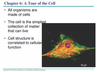
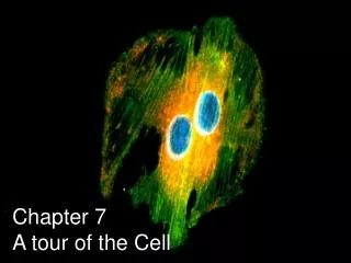
Chapter 7 A Tour of the Cell -- Part 1. TOPICS: How to study cells Eukaryotic vs Prokaryotic Nucleus and Ribosomes Endomembrane system Other membranous organelles. How do we study cells?. Know the different microscopes and their purposes: Light microscope (what we have at TPHS)

E N D
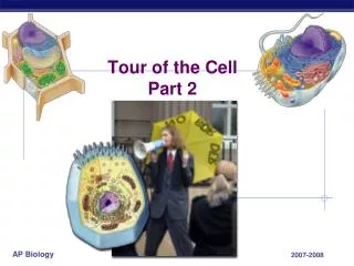
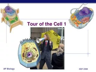
Chapter 7A Tour of the Cell -- Part 1 TOPICS: How to study cells Eukaryotic vs Prokaryotic Nucleus and Ribosomes Endomembrane system Other membranous organelles
How do we study cells? • Know the different microscopes and their purposes: • Light microscope (what we have at TPHS) • Electron microscope • SEM (scanning)—surface “scanning”, the outside (see fig. 7.2, 7.9, or 7.23 b in the book) • TEM (transmission)—”transmits” through the specimen to see inside. (see fig. 7.2, 7.13 b, or 7.18 in the book) • Figure 7.1: size range in cells
Figure 7.3cell fractionation, centrifuge What are the largest organelles/parts of a cell that fraction off into the pellet first? What are the smallest? (last)
Why are most cells so small? • THIS IS REVIEW: • Prokaryotic (“before”, “kernel” aka: nucleus) • Archaea and Bacteria, no nucleus, no membrane-bound organelles, usually much smaller, figure 7.4, they have: cytoplasm, cytosol, 1 circular chromosome, plasma membrane, cell wall, ribosomes, nucleiod. • Eukaryotic (“true”, “kernel”) • Membrane-bound nucleus and other organelles, compartmentalized cells, animals, plants, fungi, protists. These cells are larger due to compartmentalization.
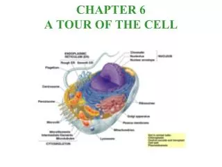
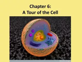
Figures on pages 108 – 109. • Know the names, identify the structures and know the functions of all of the organelles listed on these diagrams. • Know major differences between the typical animal and typical plant cells. • Animals: centrioles, • Plants: (plastids) chloroplasts, cell wall, large central vacuole, tonoplast
The Nucleus and Ribosomes • Nucleus: contains most of the genes that control a Eukaryotic cell. • Nuclear envelope/nuclear lamina: porous (why?) and double membrane • Chromatin (46)/ tightly coiled chromosomes (46), genes • Nucleolus, synthesizes ribosomes, in the nucleus • Ribosomes: site of protein synthesis • Free ribosomes=for the production of proteins to be used in the cytosol and attached ribosomes =on the ER for the production of proteins which are packaged or exported via the ER system.
Pores • Lamina • Outer andinner mem-branes • Nucleolus • Ribosomes • Attachment site of ER
Endomembrane system • Definition:all of these structures have interchangeable membranes, they are all made of a phospholipid bilayer and are fusible with one another. • Includes: nuclear envelope (lamina) ER Golgi apparatus lysosomes vacuoles plasma membrane See Figures 7.14, 7.16, 8.7 Why aren’t mitochondria and chloroplasts in this group?
The endomembrane system’s interconnectedness. All membranes are the same bilayer.
Endoplasmic Reticulum • “Network” of membranes “within the cytoplasm” • (compartmentalization: cisternal space) • Rough ER: network attached to the nucleus. Example proteins which are made from the attached ribosomes and then “shipped” via the ER: insulin, glycoproteins, transport vesicles. • Smooth ER: conducts diverse processes: synthesizes Lipids, detoxifies drugs, metabolizes carbos.: ex: glycogen hydrolysis (breakdown) in the liver.
Golgi Apparatus / Bodies • Modification and sorting of products from the ER. • Secretion organelle • Flattened sacs (cisternae), cis and trans faces, (“receiving” and “shipping/transport” sides of the golgi apparatus) • Fusion of membranes (fig 7.14, 7.16, & 8.7) is possible since the ER and the Golgi are both of the endomembrane system. • Some vesicles have external “identification” molecules *see fig. 8.7
Fig. 7.14 Notice how the vesicle from the ER fuses with the cis side of the golgi and then the trans side of the golgi fuses with a food vacuole to deliver digestive enzymes.
Lysosomes Fig. 7.14 • Contains hydrolytic enzymes which digest macromolecules and recycle materials from the cell. (see fig 7.14) • Usually maintains pH of 5 (acid)
Vacuoles • Many types: • Food vacuoles (in all cells) • Contractile vacuoles, in protists like the paramecium (*fig 8.12) for osmoregulation (water regulation). • Plants: large central vacuole (tonoplast)
Mitochondria • Energy transformation (from glucose to ATP) • Cellular respiration and ATP generation • Contain a small amount of their own DNA (semiautonomous) (Ch. 28), not of the endomembrane system. • Double phospholipid bilayer. Cristae, large surface area. Inner membrane and Matrix. • * More to come… stay tuned for Chapters 9-10 *
Chloroplasts (a type of plastid) • Energy transformations (sun energy to ATP) • Photosynthesis (CO2 and H2O to Glucose) • Synthesize organic molecules from carbon dioxide and water. • Contain a small amount of their own DNA (semiautonomous) (Ch 28) • Double membrane, thylakoids, grana, stroma fluid
Know the parts:Stroma = fluidthylakoid membranes = are chlorophyll rich grana = stacks of many thylakoids, site of light reactions (because of chlorophyll.)
Peroxisomes • Consume deadly free oxygen within the cell, transport it to mitochondria. • Enzymes transfer hydrogen to oxygen, producing hydrogen peroxide (H2O2) • H2O2 is also toxic to a cell, and an enzyme made by the peroxisome can break down H2O2