Download
1 / 1
10 likes | 118 Views
Supplementary Figure 1. Primary cancer. Tumor Cells in Ascites. Surgery. Paracentesis. Biospecimen acquisition. Biospecimen acquisition. Transport to Pathology Lab. Refrigeration. 16-24 hours. 30 minutes. Isolation of Cancer Cells. 4-8 hours. 2-4 days. Frozen sample.

E N D
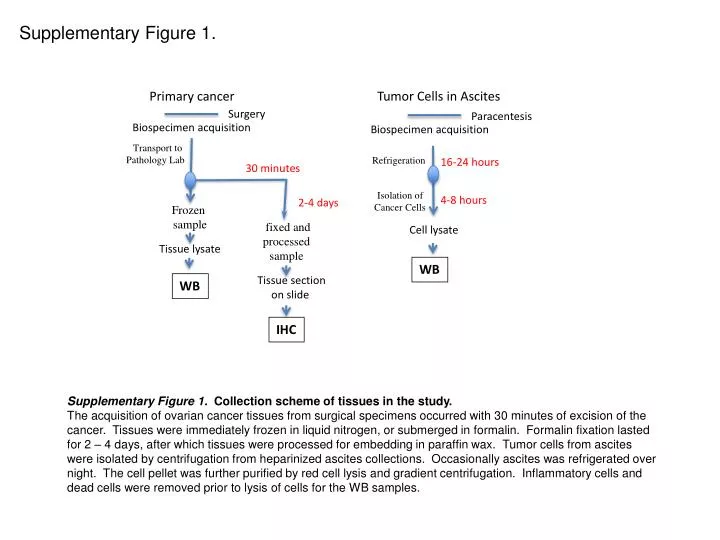
Supplementary Figure 1. Primary cancer Tumor Cells in Ascites Surgery Paracentesis Biospecimen acquisition Biospecimen acquisition Transport to Pathology Lab Refrigeration 16-24 hours 30 minutes Isolation of Cancer Cells 4-8 hours 2-4 days Frozen sample fixed and processed sample Cell lysate Tissue lysate WB Tissue section on slide WB IHC Supplementary Figure 1. Collection scheme of tissues in the study. The acquisition of ovarian cancer tissues from surgical specimens occurred with 30 minutes of excision of the cancer. Tissues were immediately frozen in liquid nitrogen, or submerged in formalin. Formalin fixation lasted for 2 – 4 days, after which tissues were processed for embedding in paraffin wax. Tumor cells from ascites were isolated by centrifugation from heparinized ascites collections. Occasionally ascites was refrigerated over night. The cell pellet was further purified by red cell lysis and gradient centrifugation. Inflammatory cells and dead cells were removed prior to lysis of cells for the WB samples.