Download
1 / 31
310 likes | 433 Views
Current UK legislation and guidelines for radiation protection of patients and staff. Prepared by: Dr D. Mladenova.

E N D
Current UK legislation and guidelines for radiation protection of patients and staff Prepared by: Dr D. Mladenova
General principles of the International Commission on Radiological Protection (ICRP) 1.No practice shall be adopted unless its introduction produces positive net benefit (Justification)2.All exposures shall be kept as low as reasonably practicable (ALARP)
Principles of ICRP- cont’d Taking economic and social factors into account( Optimization) • The dose equivalent to individuals shall not exceed the limits recommended by the ICRP (Limitation)
Regulations for intraoral radiography • Tube voltage should not be lower than 50 kV preferably 70-90 kV • Beam diameter should nor exceed 60 mm • Rectangular collimation should be used
Intraoral radiography-cont’d • Total beam filtration ( inherent and added ) • -1.5 mm aluminium disc for sets operating bellow 70 kV - 2.5 mm aluminium for sets operating above 70 kV
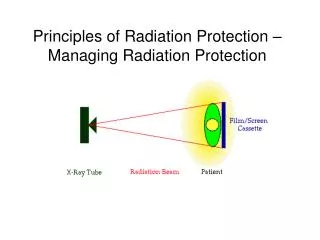
Intraoral radiography-cont’d • The focal spot should be marked on the outer casting of the tubehead • Focal spot to skin distance ( FSD) should be at least 100 mm for sets operating below 60 kV and 200 mm for sets operating above 60 kV.
Intraoral radiography-cont’d • Film speed controls and finely adjustable exposure time settings should be provided • The fastest film available( E or F speed) that will produce satisfactory diagnostic images should be used
Panoramic radiography • Equipment should have a range of tube potential settings, preferably 60-90 kV. • The beam height at the receiving slit of cassette holder should not be greater than
Panoramic radiography-cont’d • greater than the film in use (normally 125 mm or 150 mm). • The width of the beam should not be greater than 5 mm
Panoramic radiography-cont’d • Equipment should be provided with adequate patient-positioning aids incorporating light beam markers • New equipment should provide facilities for field-limitation techniques
Cephalometric radiography • Equipment must be able to ensure the precise alignment of X-ray beam, cassette and patient • The beam should be collimated to include only the diagnostically relevant area
Cephalometric radiography-cont’d • To facilitate the imaging of the soft tissues, an aluminium wedge filter should be provided at the X-ray tube
All equipment • Should have a light on the control panel to show that the mains supply is switched on • Should be fitted with a light and audible warnings that gives a clear and visible indication to the operator that an exposure is taking place
All equipment-cont’d • Exposure switches (timers) should only function while continuous pressure is maintained on the switch and terminate if pressure is released • Exposure switches should be positioned so that the operator can
All equipment-cont’d • remain outside the controlled area and at least 2 m from the X-ray tube and patient
Justification -the availability and/or findings of previous radiographs - the specific objectives of the exposure in relation to the history and examination of the patient
Justification-cont’d • the total potential diagnostic benefit to the patient - the radiation risk associated with the radiographic examination
Justification-cont’d • the efficacy, benefits and risks of alternative techniques having the same objective but no or less exposure to ionizing radiation
Lead protection • There is no justification for the routine use of lead aprons for the routine use of lead aprons for patients in dental radiography • Thyroid collar to be used only in maxillary occlusal radiography
Lead protection-cont’d • Lead aprons do not protect against scattered radiation for adult who support a patient during exposure • Lead aprons should not be folded
Specific requirements for pregnant women • Only radiographs that are absolutely necessary are taken • ALARP and the patient is given the option to delay the radiography
Selection criteria in Dental Radiography 1998 • No radiographs should be taken without a history and clinical examination • New patient • Child (primary and mixed dentition)- panoramic and bitewings radiographs
Criteria for radiographs-cont’d • Adult (dentate patient)-patient-specific radiographs depending on clinical examination • Edentulous patients- panoramic and/or periapical radiographs in selected areas
Dose limitations and annual dose • Patients • Radiation workers ( classified and non- classified) • General public
Radiographic investigations for patients • Examinations directly associated with illness • Systematic examinations • Examinations for occupational, medico-legal or insurance purposes • Examinations for medical research
Annual dose limits-cont’d • Non-classified workers 6 mSv • General public 1 mSv • Dose Constraints • non-classified workers 1 mSv
Annual dose limits-cont’d • For employee not directly Involved with radiography and General public 0.3 mSv • Pregnant staff member 1 mSv
Source of radiation to dentist and their staff • The primary beam, if they stands in its path • Scattered radiation from the patients • Radiation leakage form the tubehead
Protective measures • Personal monitoring is recommended for practice exceeding 100 intraoral or 50 panoramic film • Staff should stand outside the controlled area and not in the line of the primary beam
Protective measures-cont’d • Safe use of equipment • Safe use of radiographic techniques
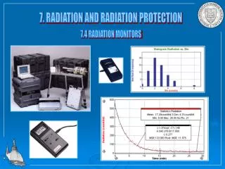
Main methods of monitoring and measuring radiation dose • Film badges • Thermoluminescent dosemeters • Ionizing chambers