Download
1 / 47
620 likes | 1.09k Views
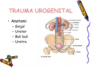
Urogenital Embryology. Robert Coleman 2008. Kidney Development. 3 embryonic kidneys, in order of appearance pronephros mesonephros metanephros all three kidneys develop from the intermediate mesoderm first two regress in utero, and the third becomes the permanent kidney. Neural tube

E N D
Urogenital Embryology Robert Coleman 2008
Kidney Development • 3 embryonic kidneys, in order of appearance • pronephros • mesonephros • metanephros • all three kidneys develop from the intermediate mesoderm • first two regress in utero, and the third becomes the permanent kidney
Neural tube • Amniotic cavity • Paraxial mesoderm • Intermediate mesoderm • Yolk sac (umbilical vesicle) • Notochord • Aorta • Intraembryonic coelom Towards the end of week3: the intermediate mesoderm moves ventrally and loses its connection (black arrow) to the somites and the lateral mesoderm.
Pronephros • transitory, nonfunctional kidney • first seen late in the 3rd week, completely degenerates by the start of the 5th week • develop as 5-7 paired segments in the region of the future neck and thorax • development of pronephric tubules starts at the cranial end of the nephrogenic cord and progresses caudally • as each tubule matures, it immediately begins to degenerate along with the segment of the nephric duct to which the tubules are attached
Mesonephros • also transient • serves as an excretory organ for the embryo while the metanephros begins its development • development of the mesonephric (wolffian) ducts precedes the development of the mesonephric tubules • soon after the appearance of the mesonephric ducts during the 4th week, mesonephric vesicles begin to form • differentiation progresses caudally and results in the formation of 40 to 42 pairs of mesonephric tubules • cranially located tubules start to degenerate at about the 5th week
Mesonephros • almost completely disappear by the 4th month • certain elements are retained as part of the reproductive tract: • ♂: epididymis and vas deferens are formed from the mesonephric (wolffian) ducts cranial mesonephric tubules become the efferent ductules of the testis • ♀: remnants of cranial and caudal mesonephric tubules form small, nonfunctional mesosalpingeal structures called the epoöphoron and the paroöphoron (and Gartner’s ducts in the vaginal vestibule) • mesonephric tubules differentiate into excretory units that resemble an abbreviated version of an adult nephron
Metanephros • definitive kidney • forms in the sacral region • ureteric buds sprout from the distal portion of the mesonephric duct and come in contact with the blastema of metanephric mesenchyme at about the 28th day • ureteric bud penetrates a condensing metanephric mesenchyme and begins to divide dichotomously; exert reciprocal inductive effects • nephrogenesis is completed before birth
Development of Nephrons/Collecting System • the nephron (glomerulus, proximal tubule, loop of Henle, and distal tubule) is thought to derive from the metanephric mesenchyme • the collecting system (collecting ducts, calyces, pelvis, and ureter) formed from the ureteric bud • older, more differentiated nephrons are located in the inner part of the kidney near the juxtamedullary region and newer, less differentiated nephrons are found at the periphery
Development of the Collecting System collecting system (collecting ducts, calyces, pelvis, and ureter) formed from the ureteric bud bifurcation of the ureteric bud determines the eventual pelvicalyceal patterns and their corresponding renal lobules first few divisions give rise to renal pelvis, calyces, and collecting ducts by 6th week, the ureteric bud has bifurcated at least four times, yielding 16 branches by 7th week, the next four generations of branches also fuse, forming the minor calyces by the 32nd week, approximately 11 additional generations of bifurcation have resulted in 1-3 million branches, which will become the collecting duct tubules
Renal Ascent 6th to 9th weeks: kidneys ascend to a lumbar site just below adrenals precise mechanism responsible unknown speculated that the differential growth of the lumbar and sacral regions of the embryo plays a role as the kidneys migrate, they are vascularized by a succession of transient aortic sprouts that arise at progressively higher levels final pair forms in the upper lumbar region and becomes the definitive renal arteries • occasionally, a more inferior pair of arteries persists as accessory lower pole arteries • when the kidney fails to ascend properly, its location becomes ectopic
ADRENAL DEVELOPMENT • cortex develops from mesoderm • medulla develops from neuroectoderm • Cortex • Begins to develop in week 5 • mesothelial cells between root of the mesentery and gonad proliferate and invade the mesenchyme: form the fetal cortex • second migration of cells forms the definitive cortex • intimate relationship with the developing gonad, kidney, and adrenal generally explains the finding of ectopic or aberrant adrenal tissue • usually associated with the kidney • Microscopic: mature adrenal cortex constitutes 90% of the gland and is divided into three zones • zona glomerulosa, zona fasciculata, and zona reticularis • zonation complete by 18 months • adult configuration is not reached until age 10-12 years • glomerulosa site of aldosterone production • fasciculata and reticularis form a single functional zone that produces glucocorticoids, androgens, and estrogens
ADRENAL DEVELOPMENT • Medulla • derived from cells of the neural crest that migrate at the seventh week to form collections, which enter the fetal cortex, leaving nodules of neuroblasts scattered throughout the cortex • by 20th week, there is a primitive medulla, but the distinct medulla is not present until atrophy of the fetal cortex • produces both noradrenaline and adrenaline, facilitated in the presence of glucocorticoids
BLADDER AND URETER DEVELOPMENT Formation of the Urogenital Sinus • 4th week, the neural tube and the tail grow dorsally and caudally, projecting itself over the cloacal membrane • 5th to 6th weeks: cloaca partitions into an anterior urogenital sinus and a posterior anorectal canal; occurs by the fusion of two lateral ridges of the cloacal wall and by a descending urorectal septum
Formation of the Urogenital Sinus • mesonephric (wolffian) duct fuses with the cloaca by the 24th day and remains with the urogenital sinus during the cloacal separation • entrance of the mesonephric duct into the primitive urogenital sinus serves as a landmark distinguishing the cephalad vesicourethral canal from the caudal urogenital sinus • vesicourethral canal gives rise to bladder and pelvic urethra • caudal urogenital sinus forms phallic urethra for males and distal vaginal vestibule for females
Formation of the Trigone • by day 33, the common excretory ducts (the portion of the mesonephric ducts distal to the origin of the ureteric buds) dilate and become absorbed into the urogenital sinus • right and left common excretory ducts fuse in midline, forming the primitive trigone • the ureteric orifice exstrophies and evaginates into the bladder by day 37 and begins to migrate in a cranial and lateral direction within the floor of the bladder • During this process, the nephric duct orifice diverges away from the ureteric orifice and migrates caudally, flanking the paramesonephric (müllerian) duct at the level of the urogenital sinus. This is the site of the future verumontanum in males and vaginal canal in females
Ectopic ureter & Weigert-Meyer rule • Upper pole UO • rotates posteriorly relative to the lower pole orifice and assumes a more caudal and medial position • may result from ureteric bud arising too high on the mesonephric duct • may drain at the bladder neck and verumontanum, or it may remain connected to the mesonephric duct derivatives • in females, the ectopic upper pole ureter may insert into the remnants of the mesonephric (wolffian) ducts or into the vaginal vestibule
Weigert-Meyer rule • Lower pole UO • assumes a more lateral position • may result from ureteric bud arising too low on the mesonephric duct • causing premature incorporation and migration within the developing bladder • VUR is more likely to occur because of an inadequate intramural tunnel
Development of the Ureter • begins as a simple cuboidal epithelial tube, surrounded by loose mesenchymal cells, which acquires a complete lumen at 28 days • undergoes a transient luminal obstruction between 37 and 40 days and subsequently recanalizes • recanalization process begins in the midureter and extends in a bidirectional manner both cranially and caudally • urine production is followed by proliferative changes in the ureteral epithelium (bilaminar by 10 weeks of gestation) • epithelium attains a transitional configuration by 14 weeks • smooth muscle differentiation is first detected in the subserosal region of the bladder dome and extends toward the bladder neck and urethra, whereas smooth muscle differentiation of the ureter occurs later within the subepithelial region in the ureterovesical junction, ascending toward the intrarenal collecting system
Development of the Bladder and Continence Mechanisms • Embryologically, bladder composed of two regions: • bladder body • derived from the endoderm-lined vesicourethral canal and the surrounding mesenchyme • trigone • develops from the incorporation of the common excretory ducts (the portion of mesonephric ducts caudal to the origin of the ureteric bud) into the base of the developing bladder
GENITAL DEVELOPMENT A The site of the primordial germ cell origin in the wall of the yolk sac in a 3-week-old embryo. B During the 5th weekprimordial germ cells migrate from the yolk sac along the dorsal mesentery to posterior body wall near the 10th thoracic level
Formation of Genital Ridges • 5th week • migration of primordial germ cells to the posterior body wall near T10 signals the mesonephros and adjacent coelomic epithelium to proliferate and form a pair of genital ridges just medial to the developing mesonephros • 6th week • cells of the genital ridge invade the mesenchyme in the region of future gonads to form primitive sex cords • the genital ridge mesenchyme containing the primitive sex cords is divided into the cortical and medullary regions • after the 6th week they pursue different fates in male and female embryos
Paramesonephric (Müllerian) duct • begin to form during week6 just lateral to the mesonephric ducts • extend all the way from the third thoracic segment to the posterior wall of the developing urogenital sinus • caudal tips are adherent to each other as they connect with the urogenital sinus between the openings of the right and left mesonephric ducts
Development of Male Genital Structures • SRY causes cells in the medullary region of the primitive sex cords to differentiate into Sertoli cells while the cells of the cortical sex cords degenerate • otherwise (no SRY), the sex cords differentiate into ovarian follicles • week 7 • differentiating Sertoli cells organize to form the testis cords • müllerian-inhibiting substance (MIS) (glycoprotein hormone) is secreted by Sertoli cells as they begin differentiating in response to SRY • week 8-10 • MIS causes the paramesonephric (müllerian) ducts to regress rapidly, leaving remnants: • small tissue protrusion at the superior pole of the testicle, called the appendix testis • posterior expansion of the prostatic urethra, called the prostatic utricle
Development of Male Genital Structures • week 9-10 • Leydig cells differentiate from mesenchymal cells of the genital ridge in response to the SRY protein • Leydig cells produce testosterone • in early development, testosterone secretion is regulated by placental chorionic gonadotrophin, but eventually the pituitary gonadotrophins assume control of androgen production • testosterone secretion stimulates mesonephric ducts to transform into vas deferens (week 8-12) • cranial portions of the mesonephric ducts degenerate, leaving small remnant of tissue called the appendix epididymis • region of mesonephric ducts adjacent to the presumptive testis differentiate into the epididymis
Development of Male Genital Structures • week 9 • 5-12 mesonephric ducts in the region of the epididymis make contact with the sex cords of the future rete testis • mesonephric tubules do not actually establish communication with rete testis as the efferent ductules until the 3rd month
Prostate • develops from the urethra • driven by DHT • begins to develop during the 10th week as a cluster of endodermal evaginations • initially form ~5 independent groups of solid prostatic cords • 11th week, cords develop lumens and glandular acini • 13th week, testosterone rises, secretory activity begins • mesenchyme surrounding endoderm-derived prostatic acini differentiates into smooth muscle and connective tissue • as prostate develops, paired bulbourethral glands sprout from the urethra just below the prostate • like renal and bladder development, prostatic development depends on mesenchymal-epithelial interactions, but under the influence of androgens
Seminal Vesicles • develop from distal mesonephric ducts • Vas and seminal vesicle devel driven by testosterone • portion of vas distal to the developing seminal vesicle is thereafter called the ejaculatory duct
Development of Female Genital Structures • primitive sex cords do not elaborate SRY protein (no Y chromosome) and therefore do not differentiate into Sertoli cells • in the absence of Sertoli cells and SRY protein, MIS synthesis, Leydig cell differentiation, and androgen production do not occur • primitive sex cords degenerate and the mesothelium of the genital ridge forms secondary cortical sex cords • invest the primordial germ cells to form the ovarian follicles • germ cells differentiate into oogonia and enter the first meiotic division as primary oocytes • follicle cells then arrest further germ cell development until puberty • mesonephric (wolffian) ducts degen, leaving remnants: • epoöphoron and paroöphoron in the mesentery of the ovary • Gartner's duct cysts near the vaginal introitus and anterolateral vaginal wall
Development of Female Genital Structures • paramesonephric (müllerian) ducts • give rise to fallopian tubes, uterus, and upper ⅔ of vagina • distal tips adhere to each other and fuse with the posterior wall of the urogenital sinus • forms uterovaginal canal which becomes superior portion of vagina and uterus • unfused, superior portions of the paramesonephric ducts become the fallopian tubes (oviducts) • funnel-shaped superior openings of the paramesonephric ducts become the infundibula • endodermal tissue of the sinusal tubercle in the posterior urogenital sinus forms a pair of swellings called the sinovaginal bulbs (while the uterovaginal canal is forming) • gives rise to the lower third of the vagina
Development of Female Genital Structures • inferior portion of the uterovaginal canal becomes occluded transiently by the vaginal plate • elongates 3rd-5th month and subsequently becomes canalized to form inferior vaginal lumen • as vaginal plate forms, the lower end of the vagina lengthens, and comes to rest on the posterior wall of the definitive urogenital sinus (future vestibule of the vagina) during the 4th month • endodermal membrane temporarily separates vaginal lumen from cavity of definitive urogenital sinus • degenerates partially after 5th month, but remnant persists as vaginal hymen
Molecular mechanism of male and female genital development. SF1 and WT1 expression is critical for genital ridge specification. SRY and SOX9, influenced by GATA4 and Fog2, are important factors for specifying the differentiation of Sertoli cells. SF1 is also critical in the regulation of MIS and other genes involved in androgen synthesis. No specific female factors have been identified, but Wnt4 and DAX1 are expressed with unique female patterns.
Development of External Genitalia • early development similar in both sexes • week 5: • pair of swellings (cloacal folds) develop on either side of the cloacal membrane • meet just anterior to the cloacal membrane to form midline swelling called genital tubercle • cloaca divides into anterior urogenital sinus and the posterior anorectal canal • part of the cloacal folds flanking the opening of the urogenital sinus becomes the urogenital folds • part flanking the opening of the anorectal canal becomes the anal folds • new pair of swellings (labioscrotal folds) then appears on either side of the urogenital folds
Development of External Genitalia • week 6: • urogenital sinus cavity extends onto surface of genital tubercle as endoderm-lined urethral groove • becomes temporarily filled by a solid endodermal structure called the urethral plate • urethral plate disintegrates and recanalizes to form an even deeper secondary groove • genital tubercle elongates to form the phallus, and a primordium of the glans clitoris and glans penis is demarcated from the phallic shaft by a coronary sulcus • appearance of the external genital is similar in male and female embryos until the 12th week
Male external genitalia • 4th month • effects of DHT on the male external genital become readily apparent • perineal region separating the urogenital sinus from the anorectal canal begins to lengthen • labioscrotal folds fuse in the midline to form the scrotum • urethral folds fuse to enclose the penile urethra • penile urethra is completely enclosed by the 14th week
Male external genitalia • distal glanular urethra formation may occur by a combination of two separate processes • fusion of urethral folds proximally • A small region of the distal urethra in the glans is formed by the invagination of surface epithelial tag with ingrowth of ectodermal cells distally • stratified squamous lining of the fossa navicularis results from an ingrowth of surface ectoderm as far proximally as the valve of Guérin
Female external genitalia • primitive perineum does not lengthen, and the labioscrotal and urethral folds do not fuse across the midline • phallus bends inferiorly, becoming the clitoris • definitive urogenital sinus becomes the vestibule of the vagina • urethral folds become the labia minora, and the labioscrotal folds become the labia majora • external genitalia develop in a similar manner in genetic males who are deficient in 5α-reductase and therefore lack dihydrotestosterone
Male genitalia 4th month Female genitalia 4th month
Gonadal Descent • testes and the ovaries both descend from original position near the 10th thoracic level • gubernaculum • necessary for initial descent of the gonads • offers the most obvious explanation of why fetal testicle descends to scrotum while ovary does not • forms during 7th week • superior end is attached to the gonad • expanded inferior end (bulb) is attached to the fascia between the developing external and internal oblique muscles in the region of the labioscrotal folds • processus vaginalis • forms at same time as gubernaculum • slight evagination of the peritoneum which develops adjacent to the gubernacular bulb • normally degenerates but occasionally remains patent (resulting in either communicating hydrocele or indirect inguinal hernia)
Gonadal Descent • inguinal canal • caudal evagination of the abdominal wall that forms when the processus vaginalis expands inferiorly, pushing out a sock-like evagination through the abdominal wall layers • first layer encountered is transversalis fascia (lies just deep to transversus abdominis) • next picks up internal oblique (becomes cremasteric muscle of the cord) • finally picks up external oblique (becomes external spermatic fascia) • ♂: inguinal canal extends down to the scrotum and allows passage of the descending testicle • ♀: complete inguinal canal also forms but appears to play no role in genital development
Testicular Descent • 3 phases of descent • transabdominal (8-23 weeks) • transinguinal (23-28 weeks) • extracanalicular (28-31 weeks) • relative growth of the lumbar vertebral column is probably responsible for intra-abdominal descent, as position is relatively fixed by the gubernacular anchoring near the inguinal canal • remain near the internal inguinal ring between 3rd and 7th month and later pass through inguinal canal in response to renewed shortening of gubernaculums • transinguinal phase appears to be a rapid process, probably occurring within a few days • once they pass through inguinal canal, testicles remain within subserosal fascia of the processus vaginalis, through which they descend toward the scrotum • further descent from external inguinal ring to dependant portion of the scrotum may take 4-6 weeks • by 9th month most testicles have completely entered the scrotal sac • gubernaculum remains as small ligamentous band attaching inferior pole of testis to scrotal floor
Mechanism of gonadal descent. The undifferentiated gonad is initially located high in the abdomen, anchored by the cranial suspensory ligament (CSL). ♂: Insl3 (gene product of the Leydig cells) causes the swelling and enlargement of gubernaculum to pull the developing testicle toward the inguinal region, and androgens cause an involution of CSL. Because of the action of MIS, müllerian ducts regress, while androgens continue to stimulate the development of wolffian ducts into male genital ductal structures. ♀: CSL persists because of the absence of androgens, and gubernaculum remains thin because of the absence of Insl3 activity, thereby keeping the ovary well within the pelvis.
Ovarian Descent • descend and become suspended within the broad ligaments of the uterus • gubernaculum-like structure extends initially from the inferior pole of the gonad to the subcutaneous fascia of the presumptive labioscrotal folds • later penetrates abdominal wall and becomes the round ligament • gubernaculum does not shorten as in males but still causes the ovaries to descend during 3rd month (by anchoring the ovaries in the pelvis) and places them into a peritoneal fold (the broad ligament of the uterus) • translocation of ovaries appears to occur during the 7th week, when the gubernaculum becomes attached to the developing paramesonephric (müllerian) ducts • paramesonephric ducts fuse together in their caudal ends and sweep out the broad ligaments and simultaneously pull the ovaries into these peritoneal folds • inferior gubernaculum becomes the round ligament of the uterus and attaches the fascia of the labia majora to the uterus, while the superior gubernaculum becomes the ligament of the ovary, connecting the uterus to the ovary • processus vaginalis of the inguinal canal is normally obliterated (occasionally remains patent to become an indirect inguinal hernia)