Download
1 / 33
330 likes | 424 Views
Chapter 46. Fig. 46-1. Animal Reproduction. Fig. 46-2. Fig. 46-3. Sexual reproduction. Asexual reproduction. Female. Generation 1. Female. Generation 2. Male. Generation 3. Generation 4. Both are females; Parthenogenic. Fig. 46-4a. The sexual behavior the cycle of ovulation.

E N D
Chapter 46 Fig. 46-1 Animal Reproduction
Fig. 46-3 Sexual reproduction Asexual reproduction Female Generation 1 Female Generation 2 Male Generation 3 Generation 4
Both are females; Parthenogenic Fig. 46-4a
The sexual behavior the cycle of ovulation Ovary size Fig. 46-4b Ovulation Ovulation Progesterone Estradiol Hormone level Time Behavior Male-like Male-like Female Female
Fertilization Fig. 46-5 Eggs
Giant water bugs: parental protection; few offsprings Fig. 46-6
Fig. 46-7 Accessorygland Ovary Ejaculatoryduct Testis Oviduct Spermatheca Penis Vagina Vas deferens Accessorygland Seminalvesicle (a) Male honeybee (drone) (b) Female honeybee (queen)
A hermaphrodite 雌雄同體 Genitalpore (Digestive tract) Fig. 46-8 Female organs: Male organs: UterusYolk gland 3 Seminalvesicle 4 Yolk duct Sperm duct(vas deferens) 3 Oviduct 2 Ovary 1 Seminalreceptacle Vas efferens 2 Testis 1 (Excretory pore)
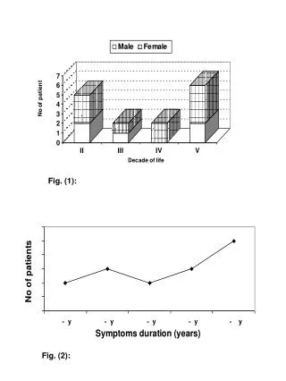
Why is sperm usage biased when female fruit flies mate twice? RESULTS Fig. 46-9 30 20 Percentage of femaleslacking sperm in spermatheca 10 0 Remated to“no-sperm”males Remated to“no-ejaculate”males Control;notremated Remated towild-typemales
Fig. 46-10a Oviduct Ovary Uterus (Urinary bladder) (Pubic bone) (Rectum) Cervix Urethra Shaft Vagina Clitoris Glans Prepuce Labia minora Labia majora Vaginal opening
Fig. 46-10b Oviduct Ovaries Follicles Corpus luteum Uterine wall Uterus Endometrium Cervix Vagina
Fig. 46-11b (Urinarybladder) (Urinaryduct) Seminal vesicle (Rectum) (Pubic bone) Vas deferens Erectiletissue Ejaculatory duct Prostate gland Urethra Penis Bulbourethral gland Glans Vas deferens EpididymisTestisScrotum Prepuce
Fig. 46-12b Epididymis Seminiferous tubule Sertoli cellnucleus Spermatogonium Primary spermatocyte Testis Cross sectionof seminiferoustubule Secondary spermatocyte Spermatids(two stages) Sperm Lumen ofseminiferous tubule
Primordial germ cell in embryo Mitotic divisions Spermatogonialstem cell 2n Fig. 46-12c Mitotic divisions Spermatogonium 2n Mitotic divisions Primary spermatocyte 2n Meiosis I n n Secondary spermatocyte Meiosis II Earlyspermatid n n n n Differentiation (Sertolicells provide nutrients) Sperm n n n n
Ovary Oogenesis Fig. 46-12e Primaryoocytewithinfollicle In embryo Growingfollicle Primordial germ cell Mitotic divisions 2n Oogonium Mitotic divisions Primary oocyte(present at birth), arrestedin prophase of meiosis I Mature follicle 2n Rupturedfollicle Completion of meiosis I and onset of meiosis II Firstpolarbody n n Secondary oocyte,arrested at metaphase of meiosisII Ovulatedsecondary oocyte Ovulation, sperm entry Completion of meiosis II Secondpolarbody Corpus luteum n Fertilized egg n Degeneratingcorpus luteum
Ovary Rupturedfollicle Primaryoocytewithinfollicle Fig. 46-12f Ovulatedsecondary oocyte Growingfollicle Corpus luteum Mature follicle Degeneratingcorpus luteum
In embryo Primordial germ cell Mitotic divisions Fig. 46-12g 2n Oogonium Mitotic divisions Primary oocyte(present at birth), arrestedin prophase of meiosis I 2n Completion of meiosis Iand onset of meiosis II Firstpolarbody n n Secondary oocyte,arrested at metaphase of meiosis II Ovulation, sperm entry Completion of meiosis II Secondpolarbody n Fertilized egg n
(a) Control by hypothalamus Inhibited by combination of estradiol and progesterone Hypothalamus – Stimulated by high levelsof estradiol + GnRH Fig. 46-14a Inhibited by low levels of estradiol Anterior pituitary – LH FSH Pituitary gonadotropinsin blood (b) LH FSH FSH and LH stimulatefollicle to grow LH surge triggersovulation Ovarian cycle (c) Corpusluteum Degeneratingcorpus luteum Growing follicle Maturingfollicle Luteal phase Ovulation Follicular phase Days | | | | | | | | 20 25 14 15 28 5 10 0
(d) Ovarian hormones in blood Peak causesLH surge Fig. 46-14b Progesterone Estradiol Ovulation Estradiol level very low Progesterone and estra-diol promote thickeningof endometrium (e) Uterine (menstrual) cycle Endometrium Menstrual flow phase Proliferative phase Secretory phase Days | | | | | | | | 0 14 15 20 25 28 5 10 Endometriosis Estrous cycle
Maternalarteries Maternalveins Fig. 46-16 Placenta Maternalportionof placenta Umbilical cord Chorionic villus,containing fetalcapillaries Fetalportion ofplacenta(chorion) Maternal bloodpools Uterus Umbilicalarteries Fetal arteriole Fetal venule Umbilicalvein Umbilical cord
Fig. 46-17 (c) 20 weeks (b) 14 weeks (a) 5 weeks
Fig. 46-17a (a) 5 weeks
Fig. 46-17b (b) 14 weeks
Fig. 46-17c (c) 20 weeks
Oxytocin Estradiol + fromovaries from fetusand mother’sposterior pituitary Fig. 46-18 Induces oxytocinreceptors on uterus Positive feedback Stimulates uterusto contract Stimulates placenta to make + Prostaglandins Stimulate morecontractionsof uterus
Placenta Umbilical cord Uterus Fig. 46-19-4 Cervix Dilation of the cervix 1 Expulsion: delivery of the infant 2 Uterus Placenta(detaching) Umbilicalcord Delivery of the placenta 3
Male Female Method Event Event Method Production ofsperm Production ofprimary oocytes Vasectomy Combination birth controlpill (or injection, patch, orvaginal ring) Fig. 46-20 Oocytedevelopmentand ovulation Sperm transportdown maleduct system Abstinence Abstinence Condom Female condom Coitusinterruptus(very highfailure rate) Spermdepositedin vagina Capture of theoocyte by theoviduct Tubal ligation Spermicides;diaphragm;cervical cap;progestin alone(as minipill,implant,or injection) Spermmovementthroughfemalereproductivetract Transportof oocyte inoviduct Meeting of sperm and oocytein oviduct Morning-afterpill; intrauterinedevice (IUD) Union of sperm and egg Implantation of blastocyst in endometrium
1. Describe oogenesis and spermatogenesis; describe three major differences between them 2. Explain how the uterine and ovarian cycles are synchronized and describe the functions of the hormones involved You should now be able to: