Download
1 / 27
290 likes | 636 Views
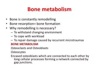
BONE METABOLISM. BONE MATRIX. Inorganic matrix – 65% Hydroxyapatite – Ca 10 (PO 4 ) 6 (OH) 2 Octacalcium phosphate - Ca 8 H 2 (PO 4 ) 6 .5H 2 O Brusite – CaHPO 4 .2H 2 O Amorphouse calcium phosphates – Ca 9 (PO 4 ) 6. Organic matrix - 20% Collagen Glycoproteins Bone specific

E N D
BONE MATRIX Inorganic matrix – 65% Hydroxyapatite – Ca10(PO4)6(OH)2 Octacalcium phosphate - Ca8H2(PO4)6.5H2O Brusite – CaHPO4.2H2O Amorphouse calcium phosphates – Ca9(PO4)6 Organic matrix -20% Collagen Glycoproteins Bone specific proteoglycans Growth factors Water – 10%
BONE CELLS Undifferentiated mesenchymal cells Osteoblasts Bone-lining cells Osteocytes Osteoclasts Ionic composition of bone mineral (mmol/g of dry fat-free bone) Anion value PO3- 4.02 CO32- 0.79 Citrate3- 0.05 Cl- 0.02 Cation value Ca2+ 6.66 Mg2+ 0.18 Na+ 0.32 K+ 0.02
Schematic representation of physiological unit of bone tissue A = vascular space B = osteocyte C = bone canaliculi D = gap junction between adjacent osteoblasts E = connective tissue
Comparison of systemic ECF (plasma) with bone ECF (electrolyte content, mmol/l) Ca Mg K Na Pi Cl Plasma 1.5 0.7 4 140 1.8 100 Bone fluid 0.48 0.4 25 125 1.8 130 Bone-lining cells Membrane which separates bone ECF and systemic ECF
Phases of bone formation • bone formation (embryonic development) • bone growth • maintenance of the skeleton – modeling and remodeling • healing of the skeleton (soft-tissue tissue trauma, fracture, response to neoplasma, infection) There is only one mechanism of bone formation and the formation of bone follows the same sequence in development, growth, and maintenance of skeleton: • within cartilage – enchondral ossification • within an organic matrix membrane – intramembranous ossification • deposition of a new bone on existing bone – appositional ossification
Diagramatic representation of the growth plate • Verical column of chondrocytes in reverse zone (chondrocytes in different phases of the cell cycle). Chondrocytes in the reverse zone are flat and inactive, give rise to the daughter chondrocytes in the proliferative zone). • In proliferative zone chondrocytes start to proliferate. • In the zone of maturation chondrocytes start to mature and give rise matrix vesicules, sites of calcification. • In the upper hyperthrophic zone chondrocytes start to enlarge and synthesize collagen type X, and produce alkaline phosphatase. Matrix vesicules are sites of the first mineral cristals. • Lower hyperthrofic zone containes degenerating apoptotis condrocytes. Proliferating mineral crystals spread in the extracellular matrix, small vessels penetrate into the matrix, osteoblasts from bone marrow deposit new bone and synthesize bone extracellular matrix.
Matrix vesicle Matrix vesicle is lying between 2 banded type II collagen fibrils. Collagen type X, the proteoglycan link protein and hyaluronic acid binding region of proteoglycan are attached to the matrix vesicles surface. The various phosphatases - alcaline phosphatase, 5-AMPase, Ca-ATPase, nucleoside triphosphatase pyrophosphohydrolase (NTP-Pase) and inorganic pyrophosphatase (PPiase) are anchored in the membrane. In the sap – LDH, matrix vesicle proteinases, carbonic anhydrase, actin and various adhesive proteins
Components of the matrix vesicle • Alcaline phosphatase • Hydrolysis of ester phosphate at the site of mineralization to produce pyrophosphate (PP) • Hydrolysis of local PP to two phosphates (Pi) • Strong binding affinity to collagen • Carbonic anhydrase • Regulates of pH inside of matrix vesicules • Removes protons as biproduct of hydroxyapatite formation • Annexins - Ca2+ion channels • Annexin V (anchorin CII) and annexin II (calpactin)
Calcification of bone matrix is under cellular control regulated by enzymes, proteins and phospoholipid membrane: Phase 1 1. Ca2+ attraction into the vesicle (Ca-binding phospholipids and Ca2+ chanels) 2. Accumulation of PO43- - action of alcaline phosphatase 3. Precipitation of calcium phosphate (noncrystaline amophous) Phase 2 1. Matrix vesicle membrane breakdown (hydrolytic action of phospholipases and proteinases) 2. Extravesicular hydroxyapatite crystal formation and proliferation 3. Itself sustaining process (constant homeostatic supplied level of Ca2+ and PO43-)
Matrix of hyperthrofic cartilage contains typical cartilage proteoglycans and collagen type II. During nucleation phase matrix get to shrink, proteoglycans aggregate. Ca2+ from adjacent matrix withdrawn into hyperthrophic cartilage matrix. Chondrocytes and matrix vesicule produce alkaline phosphatase (production of free phosphates for calcification). Mineral crystals of hydroxyapatite are formed and mineralization continues.
Phases of enchondral ossification • hypertrophy of chondroblasts • calcification of cartilage extracellular matrix • penetration of blood capillaries • differentiation of osteoblasts and osteoclasts • osteoid formation • osteoid calcification • Remodeling of new bone • bone resorption • synthesis and secretion of the bone matrix • calcification of newly formed bone lamellae
Bone resorption and bone formation are not separated, independently regulated process. Osteoblast and osteoclasts belong to structure, known as a basic multicellular unit (BMU). Osteoclasts are responsible to bone resorption Proton pupm pH 7 pH 4 Lysosomal enzymes Acid phosphatase Collagenase Glycosidase Sulphatase Catepsins Osteoblasts are responsible to organic extracellular matrix formation.
Coupling Sequential coordination between osteoclast-mediated resorption and osteoblast-mediated formation (activation-resorption-formation- ARF) In the young healthy adult resorption and formation are balanced such that bone mass remains constant. Pathological states developed when resorption and formation are unbalaced (excessive bone loss or excessive bone resorption can occur).
Bone resoption and new formation in bone remodeling unit (BMU )
Bone resoption and new formation in the BMU http://www.roche.com/pages/facets/11/ostedefe.htm
Components which are responsible to mineralization Collagen type I Bone glycoproteins : bone sialoprotein, osteonectin, osteopontin Bone phosphoproteins Small proteoglycans Growth factors
Hormones which regulate bone metabolism Systemic hormones of calcium homeostatic system: (normal physiological level of Ca2+ is 8.5 – 10.6 mg/100 ml) Parathyroide hormone (PTH) – bone resorption (stimulation of monocytes to transform to osteoclasts). Calcitonine – bone formation (inhibition of monocyte to transform to osteoclasts). 1,25-dihydroxycholecalciferol (1,25-(OH)2 vitamin D3) – bone formation (activation of osteoblasts to synthesize collagen, promote mineralization).
Calcium homeostasis • Released by low plasma calcium. • Stimulates bone resorption. • Prevents calcium excretion by kidneys. • Stimulates calcitriol synthesis. • 25-hydroxylation in liver • 1-hydroxylation in kidney • Stimulates bone resorption. • Stimulates intestinal calcium absorption. • Is released by high plasma calcium. • Acts on bone osteoclasts to reduce bone resorption. • Net result of its action is a decline in plasma calcium & phosphate. Parathyroid hormone (parathyroid) Calcitriol (1,25-diOH-Vit. D) (Vit. D in diet) Calcitonin (thyroid)
Other systemic hormones : Glucocortocuides – inhibition of bone formation. Growth hormone (GH) – stimulationof bone formation through somatomedins (growth factors IGF-1 and IGF-2). Insulin– stimulation of synthetic activity of osteoblasts. Thyroidhormones – stimulation of osteoclasts, activation of bone remodelation. Estrogens– inhibition of bone resorption (inhibition of osteoclastic activity through specific local factors). Catecholamines – antagonists of calcitonin. Prostaglandins – different classes of prostaglandins have different effect, which is dependent on concentration (10-9 – 10-7 mol/l stimulates synthesei of collagen, 10-6 inhibits collagen synthesis.
Local factors which regulate bone metabolism Factors which stimulate osteoblasts differentiation : Bone morphogenic factor (BMF) Platelet-derived growth factor (PDGF) Fibroblast growth factor (FGF) Insulin-like growth factor 1 (IGF-1) Transforming growth factor-b (TGF-b. Factors which regulate osteoclasts: Colony-stimulating factor (CSF) – suppression of osteoclasts development. Interferons (g-interferon) – inhibition of osteoclasts differentioation. Interleukins (IL-1, IL-3, IL-6, IL-11) – stimulates osteoclasts differentiation
Biochemical markers of bone formation • Alkaline phosphatase • isoenzyme of bone alkaline phosphatase activity in serum reflects osteoblastic activity. • Osteocalcin - also called bone gamma-carboxyglutamate (Gla) protein • small, noncollagenous protein that is specific for bone tissue and dentin • its precise function remains unknown • is synthesized predominantly by the osteoblast and is mostly incorporated into the extracellular matrix of the bone. • Procollagen I extension propeptides
Biochemical markers of bone resorption • Urinary calcium • fasting urinary calcium test performed on a morning sample and corrected for creatinine excretion (urinary calcium tests lack sensitivity - reflects not only skeletal resorption, but also intestinal absorption as well as renal tubular filtration and reabsorption of calcium) • Hydroxyproline • is found mainly in collagen and represents about 13% of the amino acid content of the molecule. • Methods for measuring hydroxyproline - photometry, fluorometry and high pressure liquid chromatography (HPLC). • Urinary pyridinium crosslinks (pyridinoline and hydroxypyridinoline).
Pyridinoline intramolecular cross-links Location of the two intramolecular cross-linking sites in collagen type I. In fibrils, one site links N-telopeptide to helix and the other, C-telopeptide. Pyridinoline cross-links are trivalent, joining two telopeptides to one triple-domain.