Download
1 / 20
380 likes | 2.35k Views
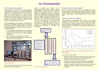
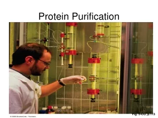
Protein Purification by Ion Exchange Chromatography. Column Chromatography. Ion Exchange Chromatography. Uses a solid matrix with either a positive or negative charge Separation is based on an equilibrium of the molecules adsorbed to the exchanger versus the elution solvent

E N D
Ion Exchange Chromatography • Uses a solid matrix with either a positive or negative charge • Separation is based on an equilibrium of the molecules adsorbed to the exchanger versus the elution solvent • Changing the ionic strength or pH of the solvent allows separation of molecules with small differences in charge
Two Types of Ion Exchangers • A cation exchanger • Bead is negative and adsorbs positively charged molecules • An anion exchanger • Bead is positive and adsorbs negatively charged molecules
If You Are Purifying a Positively Charged Molecule… • Which type of ion exchanger would you use? • Cationic • Anionic • What would be the charge on the matrix? • Positive • Negative
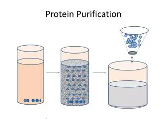
Basis for Our Ion Exchange Experiment + + - - - + - + + - - + + - + + + Sample: negatively charged protein + + + + + Beads have a positive charge
- - - - - - - Basis for Our Ion Exchange Experiment + + + + + + + + + + Sample: negatively charged protein + + + + Beads have a positive charge How can you remove the sample from the bead?
KOAc - - +- - - - - - - - Basis for Our Ion Exchange Experiment Add a salt solution (potassium acetate) Negatively-charged acetate competes for bead - + + - + - + + + + + + + Sample:Negatively charged protein + + + + Eluant: fluid/sample that is removed from column
Our Ion Exchange Experiment + - - - + + - + + + + - - + + - + + + + + Mixed Sample Negatively charged protein: GFPPositively charged protein: Cytochrome C + + + + + + + Beads have a positive charge GFP= Green Fluorescent Protein, from jellyfish, used to follow gene expression Cytochrome C= protein involved in electron transport chain
- + + - + - + - - + + - - + Our Ion Exchange Experiment + + + + + + + + + + Mixed Sample + + + +
Add 0.01 M KOAc - - - - - + + + + + Our Ion Exchange Experiment + - + + - + - + + - + + - + + + + Mixed Sample + - - + + + Cytochrome Celutes from column
Add 0.5 M KOAc - - - - - - - - - - - - - - - - - - - Our Ion Exchange Experiment + + + - + - + + + + + + + + Mixed Sample + + + + GFP elutes from column
Guide for Today’s Lab • Control flow of eluant by removing or replacing the cap on the column • Follow directions on pages 79 for • Packing the column • Separating the sample • Green Fluorescent Protein is negatively charged • Cytochrome C (yellow) is positively charged • Quantifying the sample
Guide for Today’s Lab • Follow directions on page 7 for • Packing the column (Do ALL steps listed in lab manual, only some are summarized here) • Attach column to ring stand (step 1) • Rinse with 0.01M KOAc (step 3) • Pour slurry into column (step 5) • Wash column with additional 0.01M KOAc (steps 6 & 7) DO NOT LET THE COLUMN RUN DRY
Guide for Today’s Lab • Follow directions on pages 78 for • Separating the sample • Add sample to top of column bed (steps 1 & 2) • Using 0.01 M KOAc, collect 1 ml fractions until cytochrome C (red dye) has completely eluted (steps 3-5) • Allow remaining 0.01 M KOAc to leave reservoir (step 6) • Using 0.5 M KOAc, collect 1 ml fractions until Green Fluorescent Protein (blue-green dye) has completely eluted (steps 7-10) • Measure the eluted volumes for GFP (step 11)
Guide for Today’s Lab • Follow directions on pages 89 for • Quantifying the sample • Prepare a standard curve for GFP (blue-green dye) (step 1), reading A550 for dilutions from the stock solution (step 1) • Determine the A550 value for each of the fractions containing GFP (step 2) NOTE: 5 ml samples are required for the spectrophotometer readings. You may either 1. Dilute your fractions at least 1:5 for analysis and then multiply by the dilution factor before extrapolating from the standard curve OR 2. Combine your fractions into one tube, add water if necessary to reach 5 ml and read A550. Correct for the volume when calculating the amount of GFP recovered.
Read A550THEN Use for next dilution Read A550THEN Use for next dilution H2O H2O 3 ml 3 ml 1 mg/ml 0.5 mg/ml 3 ml 3 ml Dilutions for Standard CurvePage 8 6 ml stock 1 mg/ml 0.5 mg/ml 0.25 mg/ml
Spectrophotometer Operation Set wavelengthto 550 nm Empty Pure water 100% transmittance Zero transmittance Calibration Read standards and samples at 550 nm