Download

1 / 12
250 likes | 864 Views
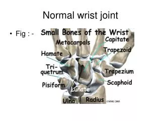
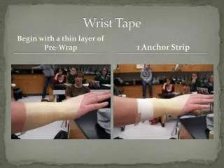
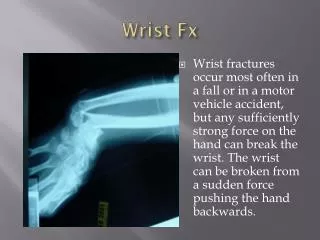
PA OBLIQES LATERAL CARPAL TUNNEL. Basic Projections. Wrist Joint. PA WRIST JOINT. Exposure Factors. Patient position Seated at end of radiographic table Apply lead shielding for Radiation safety Par t position Forearm resting on table top Axilla contact with table

E N D
PA • OBLIQES • LATERAL • CARPAL TUNNEL Basic Projections • Wrist Joint PA WRIST JOINT Exposure Factors
Patient position • Seated at end of radiographic table • Apply lead shielding for Radiation safety • Par t position • Forearm resting on table top • Axilla contact with table • Center carpals to mid cassette • Flex fingers slightly Central Ray Perpendicular Center Point To mid carpal area
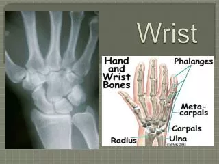
Structures shown • Mid and proximal metacarpals, carpals • Distal radius & ulna and associated joints Wrist Joint
Exposure Factors • Oblique wrist joint Patient position Seated at end of radiographic table • Apply lead shielding for Radiation safety • Part position • Forearm resting on table top with Hand in lateral position • Axilla contact with table • Center carpals to mid cassette • rotate hand medially 45 degrees
Structures shown • Distal radius &ulna • Carpals especially trapezium and Scaphoid with slight superimposition Of other carpals
Exposure Factors • Lateral wrist joint Patient position • Seated at end of radiographic table • Apply lead shielding for Radiation safety • Part position • Forearm resting on table top with • Hand and wrist in lateral position • Axilla contact with table with elbow • Flexed 90 degrees • Center carpals to mid cassette • Central Ray • Perpendicular
Center Point To wrist joint • Structures shown • Distal radius and ulna superimposed • Carpals and mid metacarpals superimposed
PA wrist With ulnar deviation ( Scaphoid) • Exposure Factors Patient position Seated at end of radiographic table Apply lead shielding for Radiation safety Part position Forearm resting on table top with Axilla contact with table & elbow Flexed 90 degrees Palmar of hand contact with table Center carpals to mid cassette Rotate wrist joint towards ulna
Central Ray Perpendicular • Center Point To Scaphoid • Structures shown Scaphoid separated from other carpals Scaphoid View
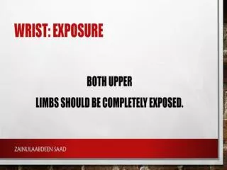
Exposure Factors • Carpal Tunnel (wrist) Patient Position Patient standing or seated at table end Ask patient to lean over and place palmar Surface of hand on cassette Part position Center palmar surface of carpals to cassette Hand and forearm near to 90 degrees angulation to cassette
Central Ray Perpendicular • Center Point Mid point of the distal forearm ( 4 cm proximal to wrist joint) Structure Shown:- Dorsal aspect of Scaphoid, lunate and trapezium Outline of the capitate and trapezium superimposed