Download
1 / 33
4.07k likes | 11.71k Views
my project presentation instagram neo_nitin if you want code of this project

E N D
Skin cancer detection using Image Processing Department Of Information Technology Rajkiya Engineering College Azamgarh, Uttar Pradesh 276201 Mentor:- Presented By:- Dr. Tauseef Ahmad Abhishek Singh(1773613003) Anurag Chaudhary(1773613011) Nitin Gaur(1773613030) Vishal Saraswat(1773613059)
Table of Content Skin Cancer (overview) Risk factors Types of skin cancer Malignant Melanoma Objective Different dataset available Block Diagram Libraries Used Model Used Workflow Testing and Comparison ROC curve and confusion matrix Result sample Reference
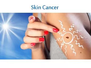
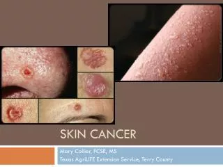
Skin Cancer • Skin cancer is the most commonly diagnosed cancer. • Skin cancers are either non-melanoma or melanoma. • Early detection and treatment can often lead to a highly favourablediagnosis • The visibility of the skin lesions increases the likelihood of early detection anddiagnosis
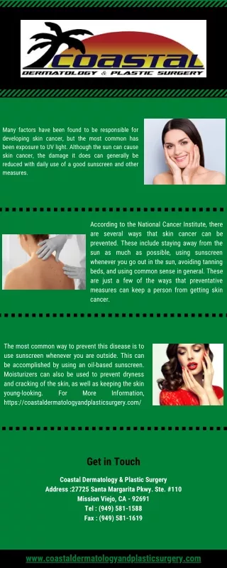
Risk Factors for SkinCancer • Damage in the ozonelayer • Fair-skinned and fair-haired people due to insufficient skinpigmentation • Prolonged exposure to sunlight. • Exposure to chemical pollutants(arsenic, nitrates, coal, tar and pitch, oils andparaffins) • History of x-ray therapy for acne orbenign lesions • Scars from severeburns • Chronic skinirritations • Immunosuppression • Geneticfactors
Types of skincancers The most common typesof skin cancer are: • Basal Cell Carcinoma(BCC) • Squamous cell (epidermoid) carcinoma(SCC) • MalignantMelanoma
MalignantMelanoma • A malignant melanoma is a cancerous neoplasm in which abnormal melanocytes are present in the epidermis and the dermis (and sometimes the subcutaneouscells). • Most lethal of all the skincancers • Most melanomas arise from cutaneous epidermal melanocytes
RISKFACTORS • The cause of malignant melanoma isunknown • Ultravioletrays are strongly suspected, based on indirect evidence such as • The increased incidence of melanoma in countries near theequator • Familyhistory
Objective • The objective of our work is detect to Melanoma Skin Cancer detection using image processing
GENERAL DIAGNOSIS BLOCK DIAGRAM Segmentation Data Collection Preprocessing Classification Feature Extraction
MAJOR LIBRARIES USED 1. Pandas 2. Numpy 3. Matplotlib 4. Keras 5. Tensorflow 6. Sklearn
MODEL USED 1. CNN Model 2. ResNet 50
Workflow • Flow Chart • Sequence Diagram • Data Flow Diagram
FLOW CHART SEQUENCE DIAGRAM
Preprocessing Images for malignant lesion (a) Original Image (b) Initial segmentation (c) Active countor model segmentation (d) Border of the segmentation on gray scale image (e) Colored ROI
CNN Model • The idea is to develop a simple CNN model from scratch, and evaluate the performance to set a baseline. The following steps to improve the model are: • Data augmentation: Rotations, noising, scaling to avoid overfitting • Transferred Learning: Using a pre-trained network construct some additional layer at the end to fine tuning our model. • Full training and Evaluation
rESnet 50 • ResNet-50 is a convolutional neural network that is 50 layers deep. • The ResNet-50(Residual Network) was introduced after CNN (Convolutional Neural Network). Additional layers are added to a DNN to improve accuracy and performance and are useful in solving complex problems. • Mostly in order to solve a complex problem, we stack some additional layers in the Deep Neural Networks which results in improved accuracy and performance. • ResNet-50 accuracy with 13 times fewer parameters and at 3 times speed better than rest of other model .
RESNET 50 IMPEMENTATION • SAMPLE IMAGE DATA
Comparison with other Model ResNet 50 CNN
MODEL LOSS ResNet 50 CNN
ROC CURVE CNN ResNet 50 Confusion matrix
REFERENCES • A. Bono, S. Tomatis, and C. Bartoli, The ABCD system of melanoma detection: A spectrophotometric analysis of the asymmetry,border, color, and dimension, "Cancer", vol. 85, no. 1, pp. 72–77, January 1999 • G.Argenziano, H. Soyer, S. Chimenti, R. Talamini, R. Corona, F. Sera, and M. Binder, Dermoscopy of pigmented skin lesions: Results of consensus meeting via the Internet Journal of the American Academy of Dermatology, vol. 48, pp. 679–693, 2003 • H. Iyatomi, H. Oka, M. Saito, A. Miyake, M. Kimoto, J. Yamagami, S. Kobayashi, A. Tanikawa, M. Hagiwara, K. Ogawa, G. Argenziano, H. P. Soyer, and M. Tanaka, Quantitative assessment of tumour extraction from dermoscopy images and evaluation of computer-based extraction methods for an automatic melanoma diagnostic system Melanoma Research, vol. 16, no. 2, pp. 183–190, 2006 • I. Maglogiannis and C. Doukas, Overview of advanced computer vision systems for skin lesions characterization IEEE Trans. on Information Technology in Biomedicine, vol. 13, no. 5, pp. 721–733, 2009 • D. Glotsos, et al. "A multiclassifer system for the characterization of normal, infectious, and cancerous prostate tissues employing transrectal ultrasound images,"Computer methods and programs in biomedicine, vol. 97, no. 1, pp. 53-61, 2010. • M. Celebi, H. Kingravi, B. Uddin, H. Iyatomi, Y. Aslandogan, W. Stoecker, and R. Moss, A methodological approach to the classification of dermoscopy images, Computerized Medical Imaging and Graphics, vol. 31, pp. 362–373, 2007
R. Kasmi and K. Mokrani, "Classification of malignant melanoma and benign skin lesions: implementation of automatic ABCD rule," IET Image Processing, vol.10, no.6,pp. 448-455, 2016. • https://arxiv.org/abs/1706.06969 • https://medium.com/@RaghavPrabhu/understanding-of-convolutional-neural-network-cnn-deep-learning-99760835f148 • https://www.mathworks.com/discovery/convolutional-neural-network.html • https://adeshpande3.github.io/adeshpande3.github.io/A-Beginner's-Guide-To-Understanding-Convolutional-Neural-Networks/