Download
1 / 73
750 likes | 958 Views
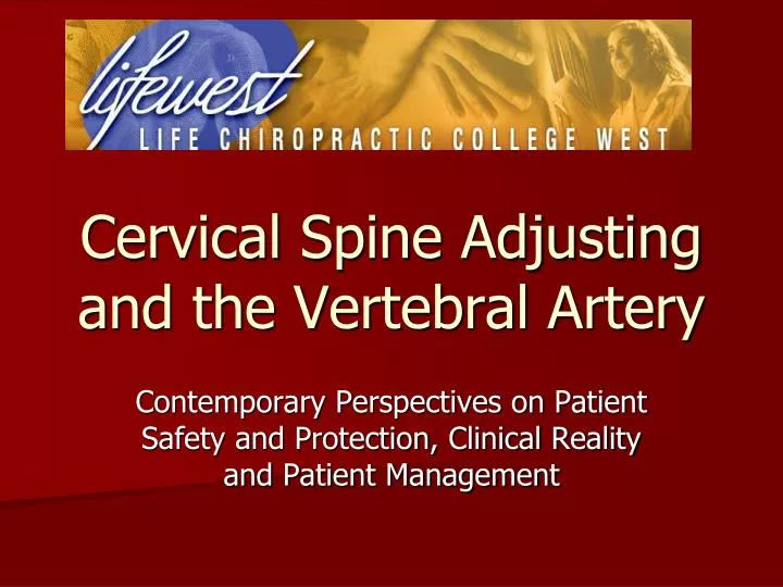
Cervical Spine Adjusting and the Vertebral Artery . Contemporary Perspectives on Patient Safety and Protection, Clinical Reality and Patient Management. Why understand this?.

E N D
Cervical Spine Adjusting and the Vertebral Artery Contemporary Perspectives on Patient Safety and Protection, Clinical Reality and Patient Management
Why understand this? Currently the single most important issue related to the practice of chiropractic from a public safety issue standpoint is associated with vertebral artery related matters. Similarly, a key issue from a public relations perspective is related to the practice of chiropractic as associated with vertebral artery related matters.
Why understand this? The Lewis Inquest in Toronto, Ontario has provided a treasure trove of information related to vertebral artery issues of interest to practicing chiropractors. The recent controversy surrounding Vioxx and Accutane signals a changing public expectation with respect to health care interventions.
Today’s Outcomes To provide the practicing chiropractor with: • A review of the relevant anatomy, physiology and pathology associated with vertebral artery injuries, particularly vertebral artery dissection, to assure an understanding of the basic mechanisms involved • A review of the current demographic and incidence data, the sources of the data, and the strengths and weaknesses of the data associated with vertebral artery injury and cervical spine adjusting
Today’s Outcomes • Current thoughts on the appropriate procedures to be used before the initiation of cervical spine adjusting and the recommended procedures in the event a patient demonstrates signs of VAD before, during, or after a care encounter • Current perspectives on VAD in progress and the clinical warning signs of the patient who presents in a potentially compromised state, as well as the most appropriate response thereto
A 30,000 foot perspective! • U.S. population: 300,000,000 • Strokes in a year in the U.S.: 700,000 • VAD occurs at a rate of: 1/100,000 pop. • VADs annually in the U.S.: 3,000 • % of pop. To visit a DC in U.S.: 10% • # of VAD patients likely to see a DC: 300 • 60,000 DCs in U.S., if evenly distributed then 1/200
Stroke Overview • 700,000 strokes annually • 80% are ischemic (clot related) • 20% are hemorrhagic (bleed related) • 3,000 VADs per year • 1 of every 233 strokes is related to a VAD • 75% of VAD related strokes make a good recovery • 5% of VAD related strokes result in death
Review: Anatomy, Physiology & Pathology of Vertebral Arteries 1. Gross anatomy 2. Histology 3. Mechanism of Basic Pathology: a. Injury and inflammation b. Clotting and thrombus formation c. Emboli d. Ischemia
1. Gross Anatomy Review Arterial circulation of the vertebral arteries: a. Origin b. Course c. Distribution d. Common anomalies
a. Origin of the Vertebral arteries: Arise from the Subclavian arteries bilaterally Arise proximal to the Thyrocervical trunk and distal to the Common Carotid artery Arterial Circulation
Divided into 4 segments as they ascend the cervical spine: Segment I: Rises from the Subclavian artery to the transverse foramen of C6 Course of the Vertebral Arteries
Segment II: Within the transverse foramina from C6-C2 Arterial Circulation
Segment III: From the superior of C2 foramen to the dura Arterial Circulation
Segment IV: From the dura into the cranium Arterial Circulation
Arterial Circulation c. Distribution from the Vertebral arteries: • From the Subclavian arteries, the Vertebral arteries ascend to ultimately unite and form the Basilar artery. • The Posterior Inferior Cerebellar artery (PICA) arises prior to the junction of the right and left Vertebral arteries forming the Basilar artery.
Arterial Circulation d. Common anomalies: Approximately 10% of patients have some form of anomaly in their Vertebral artery(ies). • Unilateral or bilateral absence of the Vertebral artery • Origin of the Vertebral arteries varies in 3.5% of cases • The PICAs have an extra dural origin in 5-20% of cases: arising approximately 1 cm proximal to dural penetration
Arterial Circulation Anomalies, continued: • Contralateral rotation can cause alteration in blood flow at the C1-C2 level on MRA • Compression of the Vertebral artery(ies) is seen in 5% of the population in both a neutral position and in rotation • Variations in arterial diameter (average 4.3 mm on the right, 4.7 mm on the left)
Arterial Circulation Anomalies, continued: • Segment 1 is tortuous in 39% of specimens • 7% of Vertebral arteries cannot be imaged with sonography due to the depth of the tissue • A change in excess of 56% is needed to detect alterations using Doppler imaging
The Vertebral arteries are comparable in size and design to the renal arteries or some of the smaller coronary arteries. They exhibit the typical 3 layer pattern from inside out: tunica intima, tunica media and tunica adventitia. 2. Histology of Blood Vessels Adventitia Media Intima
3. Mechanism of Basic Pathology a. Injury and Inflammation Arteriopathy may arise from conditions such as: • Marfan’s Disease • Ehler Danlos Syndrome-types IV and VI • Autosomal Dominant Polycystic Kidney Disease • Osteogenesis Imperfecta Type I (yielding cystic medial degeneration) • Fibromuscular Hyperplasia
3. Mechanism of Basic Pathology b. Clotting and thrombus formation • Arterial damage, particularly involving the tunica intima, will yield the start of increased localized clotting and thereby thrombus formation. • Arterial flow changes can result from histological changes as well as from mechanical changes in the vessel.
3. Mechanism of Basic Pathology Intimal dissection with blood flow beneath the intima and associated thrombus formation
3. Mechanism of Basic Pathology c. Emboli • Emboli present in three primary forms: • Liquid—amniotic fluid • Solid—excessive clotting • Gaseous—Caisson’s Disease • The thrombus at the site of arterial damage is invariably the source of emboli yielding ischemic stroke from the Vertebral artery.
3. Mechanism of Basic Pathology d. Ischemia • The degree of ischemia resultant from an embolism is the consequence of the size and location of the embolism, as well as the presence/absence of collateral circulation to the affected area.
From the Basics to the Advanced 1. Mechanisms/origins of Vertebral artery dissection (VAD) 2. Types of VAD 3. Pathophysiology of VAD 4. Sequellae of VAD
1. Mechanisms/origins of VAD The literature indicates that VAD arises spontaneously, from trivial movement, minor trauma or major trauma. The following have been cited in the literature as preceding VAD: judo, yoga, ceiling painting, nose blowing, hypertension, oral contraceptive use, sexual activity, receiving anesthesia, use of resuscitation activities, receiving a shampoo, vomiting, sneezing, adjustive or manipulative procedures.
2. Types of VAD Dissections arise from an intimal tear, yielding an intramural hematoma, which is then identified as subintimal or subadventitial. • Subintimal dissections tend to result in stenosis of the artery • Subadventitial dissections tend to result in aneurysm formation
3. Pathophysiology of VAD The root of the problem is an expanding hematoma in the wall of the Vertebral Artery as a result of: • hemorrhage of the vasa vasorum within/associated with the tunica media • development of an intimal flap in the lumen of the vessel
Subintimal vs. Subadventitial Vessel lumen Aneurysm
3. Pathophysiology of VAD The evolution of the hematoma may include the following: • It seals off, remains small and is largely asymptomatic • An expanding hematoma of a subintimal nature occludes the vessel, yielding ischemia and subsequent infarction • A lesion of a subadventitial nature yields an aneurysm that is prone to rupture through the adventitia, yielding a subdural hematoma
3. Pathophysiology of VAD The consequences of an intimal disruption may include: • alteration of normal hemodynamics • creation of a thrombogenic environment • formation of a thrombus • potential generation of emboli
4. Sequellae of VAD The effects of altered arterial flow through the Vertebral artery as a result of a dissection can yield: • Few or minimal symptoms • Transient ischemic attacks • Development of thrombi and emboli • Potential for ischemia and/or infarction
VAD in Literature 1. Mechanisms of origin 2. Incidence 3. Morbidity and mortality 4. Predisposing factors 5. Theorized predisposing factors 6. Predictors of VAD
Vertebral Artery Dissection 1. Mechanism of origin According to Haldeman et al. Spine 1999 Apr 15;24(8):785-94 (Based on cases reported in scientific literature): • 43% of are spontaneous in nature • 31% are associated with cervical spine manipulation • 16% from trivial trauma • 10% from major trauma
Vertebral Artery Dissection 1. Mechanism of origin According to Beaudry and Spence (The Canadian Journal of Neurological Sciences, V. 30, No. 4, November 2003, pp. 320-304) • The most common cause of traumatic Vertebrobasilar ischemia is motor vehicle accidents. • Of 80 cases that presented over 20 years to a single neurovascular practice, 70 were related to MVAs, 5 to industrial injuries, 5 associated with chiropractic. Consideration was offered that some of the cases that were related to chiropractors were also involved in MVAs, further confounding the matter.
Vertebral Artery Dissection 2. Incidence of VAD (Schievink, NEJM 3/22/01) • Approximately 3,000 of the 700,000 reported strokes yearly are VADs. • Dissections account for 10-25% of all ischemic strokes in young or middle aged persons. • VAD and CAD account for 2.6 per 100,000 strokes. • Female to male ratio: 3:1 (disputed) • Average age: VAD—40, CAD—47 (disputed)
Vertebral Artery Dissection 2. Incidence of VAD—Reported Numbers • 1 in 5,000 adjustments cause a stroke (Norris, SPONTADS, unpublished) • 1 in 20,000 adjustments cause a stroke (Vickers, BMJ, 1999) • 1.3 in 100,000 patients (Rothwell, Stroke, 2001) • 1 in 1 million adjustments (Hosek et al, JAMA, 1981) • 1 in 2 million adjustments (Klougart et al, JMPT, 1996) • 1 in 5.85 million cervical spine adjustments (Carey et al, CMAJ, 2001)
Vertebral Artery Dissection 2. Incidence of VAD—Discussion • The Rothwell data involves all patients who experienced a stroke within 7 days of a chiropractic office visit • The Carey data reflects claims filed for a stroke following chiropractic care • It is likely that among the Rothwell data there were unrelated strokes and among the Carey data there were unreported claims • Safe number: 1/1-2 million adjustments
Vertebral Artery Dissection 4. Predisposing factors • Aforementioned heritable conditions • “approximately 5 percent of patients with spontaneous dissection of the carotid or vertebral artery have at least one family member who has had a spontaneous dissection of the aorta or its main branches.” (Schievink, NEJM 2001)
Vertebral Artery Dissection 5. Theorized predisposing factors • Migraine—One case-control study in 1989 suggested that migraine was a risk factor for cervical artery dissection (D’Anglejan, Headache, 1989) • Hyperhomocysteinemia—reported by Pezzini, J Neurology, 2002 • Previous respiratory infection together with other neurological symptoms
Vertebral Artery Dissection 6. Predictors of VAD “Thus, given the current state of the literature, it is impossible to advise patients or physicians about how to avoid vertebrobasilar artery dissection when considering cervical manipulation or about specific sports or exercises that result in neck movement or trauma.” (Haldeman et al, Spine 1999)
Clinical Pearl Number One Current thinking holds that the majority of patients who develop frank symptoms of a vertebral artery dissection following chiropractic care were in the process of dissection when they presented for care.
In Support of this Idea • Did the SMT Practitioner Cause the Arterial Injury? Terrett, Chiropractic Journal of Australia, Vol. 32, No. 3, 9/2003, pp. 99-110 • Manipulation of the Neck and Stroke: time for more rigorous evidence. Breene, Medical Journal of Australia, Vol. 176, 15 Apr 2002, pp.364-365 • Spinal manipulative therapy is an independent risk factor for vertebral artery dissection. Smith, Neurology, Vol. 60, pp. 1424-1428
Against this Idea • Spinal Manipulative Therapy is an Independent Risk Factor for Vertebral Artery Dissection. Smith, Neurology, 2003, Vol. 60, pp. 1424-1428
For the Practicing DC • Pre-adjusting screening tests • Pre and post adjustment indicators of VAD • Abnormal behavior/speech • Pathognomonic complaint • Pain patterns • Important Steps to follow
Pre-adjustment screening tests? We were all taught “George’s Test”, “DeKlynes Test” and other tests for Vertebral artery competency. Regardless of what your teachers, colleagues, professional liability carriers, risk management consultants have told you about these provocative tests—DO NOT DO THEM.
Pre-adjustment screening tests • Yield an unacceptable percentage of false positives and false negatives • Offer no reliable information • May be enough to make a bad situation worse for the patient who has a VAD in progress • Were included in provocative testing that was abandoned by all of the clinic directors of U.S. chiropractic programs (which was supported by all of the presidents/deans of the schools) in March 2004 • Bottom Line: There are no reliable or safe tests that will rule out a VAD in progress and there are no tests that will identify a patient at risk for VAD.