Download
1 / 1
20 likes | 134 Views
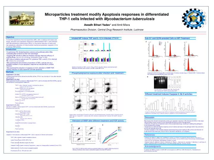
A. B. C. B. A. C. D. E. F. Spun at 1000 rpm for 5min. Removed supernatant. Incubated in dark for 15min. Re-suspended in binding buffer. PMA(20nM/ml medium). M Tb H37Ra; MOI 20; 2 hrs. Treatment (2hrs); Washing.

E N D
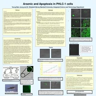
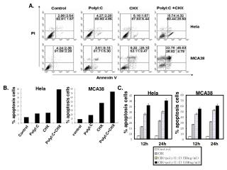
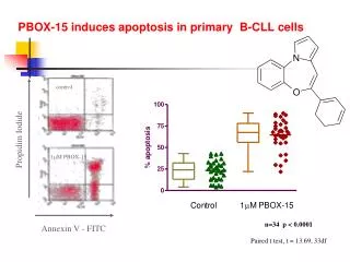
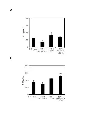
A B C B A C D E F Spun at 1000 rpm for 5min Removed supernatant Incubated in dark for 15min Re-suspended in binding buffer PMA(20nM/ml medium) M Tb H37Ra; MOI 20; 2 hrs Treatment (2hrs); Washing Microparticles treatment modify Apoptosis responses in differentiated THP-1 cells Infected with Mycobacterium tuberculosis Awadh Bihari Yadav* and AmitMisra Pharmaceutics Division, Central Drug Research Institute,Lucknow Objective Inhaled MP Induce TNF and IL-12 in Infected J774 A1. Sub-G1 and G2/M-arrested Cells on MP-Treatment To test the hypothesis that microparticles (MP), with or without anti-tubercular drugs, activate a pro-apoptotic response in differentiated THP-1 cells infected with Mycobacterium tuberculosis (M.tb.); in the terms induction of early and late apoptosis, alteration of mitochondrial membrane potential, caspase-3, 8 & 9 activity, P2R activity. A Introduction • In pulmonary TB, inhaled bacteria colonize and proliferate within AM, modulating M functions to their own advantage. • Targeting MP directly to AM via inhalation therapy improves efficacy of existing drugs. (Sen et al,PCT Int’l Pat App. 20050084455, October 16, 2003) • MP induce oxidative radicals and Th1 cytokines TNF- and IL-12 in infected M. (Sharma et al, communicated) • We examined the time kinetics of secretion of TNF- and its role as a possible mediator of a pro-apoptotic response by infected M to combat infection. • We examined the induction of caspase-3, 8 & 9, alteration of MMP, P2R activity, induction of early apoptosis and late apoptosis. Kinetics of secretion of TNF- and IL-12 by J 774 A1 cells after infection and treatment with Rifampicin+Isoniazid; either soluble or as MP or blank MP for 2h. Methods Phospthatidylserine exposure after infection and treatment A: Normal controls, B: Normal+MP, C: Infected cells, D: Infection+Soluble drugs, E: Infection+MP, F: Infection+Blank MP. Analysed using Modfit® after Kamer et al, 2005. • Experiment 1 (In vitro) • Cytokine production by the cultured murine M cell line J774 A.1 as a function of time after infection and treatment • Experiment 2 (In vitro) Early apoptosis induction analysis in differentiated TH P-1 cell line infected with M tb H37Ra, stained with annexine V and propidium iodide (PI) • THP-1 cells, (infected, treated, incubated as above) • Added 1XPBS 200 l & resuspend • Re-suspended in 1X binding buffer • Added 2 l of FITC conjugated annexine V • Spun at 1000 rpm for 5 min & supernatant removed • Added 10 l of PI • Flow cytometry • Experiment 3 (In vitro) • Alteration of MMP in differentiated THP-1 cell line infected with M tb H37Ra, stained with Rhodamine123 (Rh123) • THP-1 cell line (Monocytes) • Differentiated THP-1 cells • Infected cells • Incubated 12hrs • Added Rh123 (5mM), incubate 30min. • Washed • Flow cytometry Experiment 4 (In vitro) • Caspase-3, 8 & 9 induction in differentiated THP-1 cells in response to infection and treatment • THP-1 cells, (infected, treated, incubated as above) • Lysed (Freeze-thaw in EGTA, EDTA, Tris) • Protein estimated (Bradford reagent, Bio-Rad) • Caspase-3 assay (Lysate containing 10g protein + equal vol. of assay buffer, incubated 30 min; 37°C). • Added substrate (10 M coumarin-conjugated peptide) • Fluorescence at ex = 360 and em= 460 B M: 1Kb marker, I: Infected cells, ISD: Infected+SD, IMP: Infected+MP, IDMP: Infected+Blank MP) Different treatment Induces Caspase-3, 8& 9 activities In graph A. Arbitrary Fluorescence Units (AFU) of Caspase-3 substrate breaking, B. Specific activity of caspase-8 in cell lysate and C. Specific activity of caspase-9 in the cell lysate plotted. Scatter plots y-axis showing PI positive cells and x-axis showing annexin V positive cells. N: Normal controls, NMP: Normal+ treated MP, I: infected cells, ISD: Infected+ treated SD, IMP: infected+ MP, IDMP: infected+ Blank MP Discussion Alteration of MMP after different treatment and P2R activity • We propose that inhaled microparticles evoke a pro-apoptotic response in M infected with M tb. in two ways- • By providing a direct stimulus to the infected M through the event of phagocytosis. • Indirectly, through the anti-bacterial action of the incorporated drugs, resulting in nullifying the pathogen’s immunosuppressive strategy. • Apoptosis is a valid anti-infective response, since it denies sanctuary to the pathogen which has evolved to survive and replicate within M. • Further evaluation of the interplay between bactericidal drug action leading to Msurvival and induction of innate bactericidal responses, some of which may lead to apoptosis, is in progress. A B References • Sharma, R., Muttil, P., Yadav, A. B., and Misra, A. Uptake of inhalable microparticles affects defense responses of macrophages infected with Mycobacterium tuberculosis H37Ra. Journal of Antimicrobial Chemotherapy. (communicated) • Sen et al,PCT Int’l Pat App. 20050084455, October 16, 2003) • Kamer,I.,Sarig, R., Zaltsman,Y., Niv, H., Oberkovitz, G., Regev, L., Haimovich, G., Lerenthal, Y., Marcellus, C. and Gross, A. (2005). Proapoptotic BID Is an ATM Effector in the DNA-damage response. Cell 122: 579-591 Acknowledgments Fig. A: FSC Vs. Mitochondrial membrane potential plotted and in graph B: Mean fluorescence intensity of ethidium influx inside a cells plotted against time after ATP addition Funded by the NMITLI program of CSIR. ABY is the recipient of a SRF from CSIR. We thank A.L. Vishwakarma for performing flow cytometry.