Download
1 / 33
350 likes | 755 Views
HEMOSTATIC DISORDERS AND MASSIVE OBSTETRIC HEMORRHAGE Alexander D. Makatsariya Moscow 201 2. The most frequent causes of the postoperative coagulopathies — the initial hemostatic abnormalities. Michael DeBakey.

E N D
HEMOSTATICDISORDERS ANDMASSIVE OBSTETRIC HEMORRHAGE Alexander D. Makatsariya Moscow 2012
The most frequent causes of the postoperative coagulopathies — the initial hemostatic abnormalities. Michael DeBakey
Every year, 529 000 women in child-bearing age die from pregnancy-related complications • In 25% fatal outcome is due to bleeding • Every day, 500 women die from bleeding
Maternal mortality (per 100,000 live-born infants) Main causes
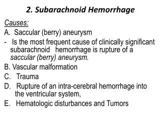
Conventional risk factors and causes Other risk factors of obstetric bleeding Uterine scar Multiparity Hydramnion, multiple pregnancy, large fetus Accelerated/prolonged labour obesity Infectious complications/Sepsis Uterine myoma Pregnancy complications: Pre-eclampsia Placenta previa Antenatal fetal death Abnormalities of placentation Chorioamnionitis Causes of massive obstetric bleeding Abruptio placenta Placenta previa Uterine hypotonia Uterine ruptures Injures of external genitals Retained placenta Amniotic fluid embolism
Severe forms of pre-eclampsia Anaphylactic syndrome Septic shock Injures of external genitals Retained placenta Antenatal fetal death (dead fetus syndrom) Abruptio placenta Placenta previa Uterine hypotonia Uterine ruptures Amniotic fluid embolism Infectious complications/Sepsis The absence of adaptive changes in the hemostasis system in pregnancy Deficiency of coagulation factors Concealed genetic bleeding diathesis Genetic and/or acquired thrombophilia Inhibitory forms of bleeding diathesis Qualitative and/or quantitative platelet's defects DIC-syndrome Inflammation SIRS Hepatic Pathology Medicamentous hemostatic disorders
According to our 25year experience obstetric bleeding can be divided into moderate and massive
Massive obstetric haemorrhage is initially coagulopathic. At the same time coagulopathy and hemostasis insufficiency have different nature
Multifactorial massive obstetric bleeding Pregnancy pathology: Placenta abruptio Severe forms of pre-eclampsia Anaphylactic syndrome Septic shock Genetic and/or acquired thrombophilia DIC-syndrome Inflammation SIRS Hepatic Pathology The absence of adaptive changes in the hemostasis system in pregnancy Massive obstetric bleeding Medicamentous hemostatic disorders Inhibitory forms of bleeding diathesis Deficiency of coagulation factors Qualitative and/or quantitative platelet's defects Concealed genetic bleeding diathesis
Risk factors of massive obstetric bleeding • The absence of adaptive changes in the hemostasis system during the pregnancy • The transition of compensated to decompensated form of DIC-syndrome • Qualitative and/or quantitative platelet's defects • Concealed hemostatic defects • Acquired hemostatic defects (the medicamentous disorders, liver diseases) e.c. Hemorrhagic diathesis Bleeding Labour Cesarean delivery Multiple abnormalities of hemostasis system Time
The absence of adaptive changes in the hemostasis system in pregnancy
Hemodynamic changes in pregnancy • Increased blood volume (for 1000-2000 ml) • Decreased total peripheral resistance • Physiologic hemodilution • Elevating the cardiac output (by more than 40-45%) • Elevating the uterine arteries blood flow from 10-15 up to 600-800 ml/min
The conversion of compensated form of DIC-syndrome to decompensated
DIC-syndrome Systemic activation of coagulation and inflammation Consumption of platelets, coagulation factors, natural anticoagulants Systemic fibrinolysis, FDPs circulation, platelet's dysfunction Systemic fibrin deposition in microvascular bloodstream Microvascular thrombosis Systemic Inflammatory Response Syndrome: hyperexpression of TF, TNF-α, Il-1, IL-6, Il-8, endothelial dysfunction Multiple organ failure syndrome Massive bleeding DIC-syndrome as a basis of pathogenesis of massive bleeding
D-dimer, fibrin degradation products (FDPs) • Inhibition of platelet's function • Inhibition of thrombin activity • Abnormal fibrin polymerization • Abnormal myometrial contractility • Abnormalities in myocardial contractile activity • Hypotonic bleeding in this situation is secondary to DIC-associated coagulopathy
Masked hemorrhagic defects • Genetic or acquired thrombocytopenia and/or thrombocytopathy • Deficiency of coagulation factors Von Willebrand Disease Hemophilia А or В Rare coagulation defects (deficiency of factors V, V и VIII, VII, X, XI, XIII, II) • Inhibitory forms(acquired antibodies to coagulation factors)
Anamnesis – the key to diagnose inherited hemostatic defects • Personal history Haemorrhage after delivery/abortion Menorrhagia Nasal (epistaxis), gingival bleeding Postoperative bleeding, including milnimal (tonsillectomy, tooth extraction) Minor formation of the bruises • Family history
Personal history – menorrhagia in patients with hemostatic defects Philipp CS, Dilley A, Miller CH et al. Platelet functional defects in women with unexplained menorrhagia. J Thromb Haemost. 2003 Mar;1(3):477-84. MillerCH, Heit JA, Kouides PA et al. Laboratory characteristics of women with menorrhagia: a multisate US tudy. J Thromb haemost 2007; 5 (Suppl 2); 187
Defects in hemostasis system in patients with coagulopathichistory Patients with history of massive obstetric bleeding, n=51 • Thrombophilia – 61% Genetic – 55% Acquired 8% Multigenic – 4% • Defects of platelets hemostasis – 31% • Von Willebrand Disease – 13% Diagnosed hemostatic defects
Changes in hemostasis system in patients with hepatic disorders: balance between the risk of bleeding and prothrombotic phenotype Bleeding disorders Thrombosis • Deficiency of factor VII • Deficiency of vitamin К • Deficiency of coagulation factors • Thrombocytopenia • Thrombocytopathy Hypofibrinolysis ↓ PAI-I ↓ α2-antiplasmin Inhibition of natural anticoagulants: ↓ Protein С ↓ Protein S ↓ Antithrombin III Increased platelets aggregation
Conclusions - The majority of obstetricians consider HYPOTONIA OF UTERUS as a main cause of obstetric hemorrhages. - But we concern that it’s true only to moderate hemorrhages which can be easily stop and are not the cause of hemorrhagic shock or fatal outcome.
The real life threatening condition represent the hemorrhages which are primarily coagulopathic and treatment of them is very difficult. IN CASES OF DIAGNOSED PRIMARILY COAGULOPATHY MASSIVE OBSTETRIC HEMORRHAGES CAN BE SUCCESSFULLY PREVENTED (BEFORE LABOR OR CESAREAN DELIVERY).
Strategy for patients with obstetric haemorrhages — Mechanical methods are not enough!!! Massive postpartum haemorrhage Uterine massage, uterotonic agents Hemostasis System examination Maintenance of blood volume Surgical methods of bleeding treatment, repairement of ruptures, ligation of the uterine blood supply, Lynch suture, embolization of uterine arteries Continuous uncontrolled haemorrhage after 8-12 U RBC Before hysterectomy → Novoseven 90 mcg/kg In the absence of effects in 20 min → repeat Novoseven 90 mcg/kg Uncontrolled bleeding - Hysterectomy. Continuous bleeding : Re-estimation of hemostasis system Initially: 4 U RBC (maintenance Нb at the level > 70 g/L) Then: 4 U RBC + 4 U FFP + 1 dose platelets + 8 U cryoprepicitate
Prophylaxis of massive obstetric bleeding begins from the evaluation ofrisk factors :
- Hemorrhagic anamnesis includingpersonal and family history menorrhagia, nasal and gingival bleeding, bruise formation, petechial hemorrhages, obstetric bleeding and others; - History of arterial and venous thromboembolism, placenta abruptio, pre-eclampsia -the evaluation of thrombophilic defects.
- diagnosis of hepatic diseases (particularly hepatitis); • revision of all received medication that influence coagulation (anticoagulants, antiaggregants and anaesthetic drugs) - the frequency of hemotransfusion (particularly in patients with inherited bleeding diathesis) for the purpose of exclusion of inhibitory forms of hemorrhages.
- In patients with thrombophilia and APS early anticoagulant prophylaxis with LWMH prevents the development of massive obstetric bleeding associated with placenta abruption, decompensated DIC, severe pre-eclampsia
- Patients with hepatic disorders, acquired and inherited hemorrhagic diathesis should be treated with clotting factors replacement therapy, fresh-frozen plasma in the second stage of labor and early postpartum. Preferably to use coagulation factors combination with natural anticoagulants
- In cases of inhibitory forms of hemorrhagic diathesis – factor replacement therapy excluding antibody-associated factors (NovoSeven in patients with Von Willebrand disease, hemophilia).