Download
1 / 21
210 likes | 398 Views
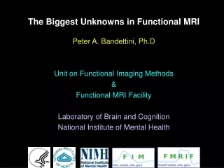
Functional MRI Facility. Core Facility Staff: Peter Bandettini, Ph.D. – Director Sean Marrett, Ph.D. – Staff Scientist Jerzy Bodurka, Ph.D. – Staff Scientist Wen-Ming Luh, Ph.D. – Staff Scientist Adam Thomas – Computer Administrator Kay Kuhns – Program Assistant

E N D
Core Facility Staff: Peter Bandettini, Ph.D. – Director Sean Marrett, Ph.D. – Staff Scientist Jerzy Bodurka, Ph.D. – Staff Scientist Wen-Ming Luh, Ph.D. – Staff Scientist Adam Thomas – Computer Administrator Kay Kuhns – Program Assistant Karen Bove-Bettis – Technologist Janet Ebron – Technologist Alda Ottley – Technologist Ellen Condon – Technologist Sahra Omar – Technologist
Scanners: “3T-1” GE 3T (June 2000) “3T-2” GE 3T (Nov 2002) “FMRIF 1.5T” GE 1.5T (Sept 2004) Currently being Cited GE 3T (Aug 2003) 1.5T 3T-1 3T-2 1.5T
Radiofrequency Coils Head “Bird-cage” coils
A B C D
Radiofrequency Coils 8-channel acquisition: GE birdcage GE 8 channel coil Nova 8 channel coil
Stimulus presentation equipment • Back projection screen 48X36in (DaLite Polacoat 100) mounted on an aluminum stand. • Sharp LCD projectors with Buhl lens • Avotec Silent Vision fiber-optic glasses for visual stimulus with integrated eye-tracking system • SMI iView system with long-range lens for video-camera based eye-tracking • Avotec Silent Scan earphones • Phone-Or Dual Channel Noise-canceling Microphone • Software and response devices • Presentation software • e-prime (biological) • Psychophysics Toolbox • SuperLab • Custom designed button response units and physiological interfaces RSB • New Devices (acquired in the last year) • EEG • Custom DLP projection (higher temporal resolution) • DLP Backprojection • Fiber-optic response systems • MRI compatible power-injector • Drug infusion pump
Pulse Sequences • BOLD imaging: • EPI-RT : General purpose BOLD imaging with real time display • epi3, epi4 : NIH EPI sequences, epi4 for use with 16 channel system • SPEP: Simultaneous perfusion and BOLD -spiral/EPI sequence with perfusion and diffusion modules and multi-echo and combined SE and GE capability • Clustered volume EPI-RT: (for auditory studies) • NIH-EPI (for use with 16 channel receiver system) • Anatomical Imaging: • MP-RAGE: T1 weighted sequence with excellent Gray/White matter contrast • standard product multi-shot sequences like: SPGR, SE, FSE etc. EPI IR-EPI Pulsed ASL (QUIPSS II) High-resolution venogram
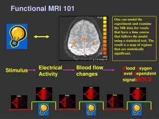
“Real – Time” fMRI motion tracking
Website: FMRIF.NIMH.NIH.GOV
3T-1 3T-2
Education / Support: • Weekly fMRI discussions (Fridays, 1pm, 10/4N230) • Bi-Monthly user meetings (First Monday every other month, 3pm, 10/4N230) • Bi-Monthly steering committee meetings (First Monday every other month, 3pm, 10/4N230) • Meetings with each PI to address needs and concerns & guide purchases • Training in scanner operation and use of subject interface devices • Consulting on paradigm design
PI Research: NINDS: Leonardo Cohen, M.D. Jeff Duyn, Ph.D. Jordan Grafman, Ph.D. Mark Hallett, Ph.D. Alan Koretsky, Ph.D. Chrsity Ludlow, Ph.D. NIAAA: Daniel Hommer, M.D. NICHD: Peter Basser, Ph.D. Allen Braun, M.D. NIMH: Peter Bandettini, Ph.D. Karen Berman, M.D. James Blair, Ph.D. Robert Cohen, M.D., Ph.D. Christian Grillon, Ph.D. Wayne Drevets, M.D. Ellen Liebenluft, M.D. Daniel Pine, M.D. Jun Shen, Ph.D. Leslie Ungerleider, Ph.D. Daniel Weinberger, M.D.
Research Protocols on FMRIF Scanners 17 32 20
2003: Up days: 3T-1: 303 3T-2: 280 Total budget (including salaries): $1,746 000.00 Cost per usage hour (only counting up days x 10 hrs day): $309 Cost per Gigabyte: $2.35
Functional MRI Papers Published at the NIH: 2000: 20 2001: 11 2002: 24 2003: 26 2004: 31 2005: 5
Ongoing Projects and Directions • More routine access to advanced subject interface devices (eye tracking, skin conductance). • Better dissemination of methods information to and across groups. • Simultaneous EEG/fMRI. • Higher resolution single shot fMRI (1.5 mm3). • More routine access to perfusion imaging methods and processing. • Better shimming techniques (to image base of brain more effectively).
Schedule so far Feb 17: Peter Bandettini: Introduction, overview March 3: (3:00 PM) Steven Schiff from the Krasnow Institute March 10: Joelle Sarlls from the University of Arizona. "Radial Data Acquisitions in Diffusion-weighted MRI" March 17: Danny Pine, Mood and Affective Disorders Program March 24: Niko Kriegeskorte (discussion: multivariate analysis and pattern classification) March 31: April 14: David Leopold April 21: Adam Thomas (discussion: slice orientation tradeoffs) April 28: Rasmus Birn May 19: Sean Marrett (discussion: fMRI of processes associated with perceptual decision making) May 26: Kyle Simmons, Laboratory of Brain and Cognition (discussion: voxel-wise uniqueness and variability of activation patterns) June 23: June 30 July 7 July 21 July 28
Potential topics/issues… Signal dropout Multivariate analysis Pooling across scanners Slice orientation DTI and fMRI integration Connectivity assessment methods High field strength issues Simultaneous measures (EEG, SCR, eye tracking, physiology) FMR-Adaptation Behavior prediction Neuronal-hemodynamic coupling Voxel based morphometry High resolution issues Perfusion imaging …?