Download
1 / 5
50 likes | 61 Views
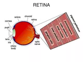
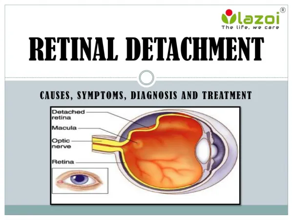
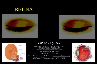
Eye is made up of Iris, Pupil, Cornea and Retina. The retina is an extremely thin tissue that lines the inside of the back of the eye. It is the light-sensitive portion of the eye. Light from the objects we are looking at, enters the eye. Cornea and the eye lens focus the light image onto the retina. Human eye works like a camera, light striking the retina causes a complex biochemical change within certain layers of the retina and this, in turn, stimulates an electrical response within other layers of the retina.

E N D
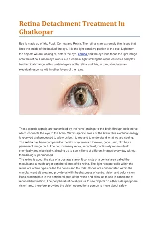
RetinaDetachmentTreatmentIn Ghatkopar EyeismadeupofIris,Pupil,Cornea andRetina.Theretinaisan extremelythin tissuethat linesthe inside ofthe backoftheeye.Itisthelight-sensitiveportion ofthe eye.Lightfrom theobjectswe arelooking at,entersthe eye.Corneaandthe eyelensfocusthe lightimage ontothe retina.Human eyeworkslike acamera,lightstriking theretinacausesa complex biochemicalchangewithincertainlayersoftheretina andthis,in turn,stimulatesan electricalresponsewithin other layersoftheretina. These electric signals are transmitted by the nerve endings to the brain through optic nerve, which connects the eye to the brain. Within specific areas of the brain, this electrical energy isreceived andprocessedtoallow usboth tosee andtounderstandwhatweareseeing. Theretinahasbeen comparedtothefilmofa camera.However,onceused,film hasa permanentimage onit.Theneurosensoryretina,incontrast,continuallyrenewsitself chemicallyand electrically,allowing ustoseemillions ofdifferentimageseverydaywithout thembeingsuperimposed. Theretinaisabout thesizeofapostagestamp.Itconsists ofa centralareacalledthe maculaand amuchlargerperipheralarea oftheretina.Thelightreceptorcellswithin the retinaareoftwo typescalledthe conesand therods.Conesareconcentratedwithin the macular (central)area andprovideuswith thesharpness ofcentralvisionandcolorvision. Rodspredominateintheperipheralareaoftheretina andallowusto seeinconditionsof reduced illumination.Theperipheralretina allowsustoseeobjectson eitherside (peripheral vision) and,therefore,providesthe visionneeded fora person tomoveaboutsafely.
RetinalDetachment • Retinaldetachmentoccurswhen theretinabecomesseparated from the nervetissuesand blood supply underneath it. While painless, visually this has a clouding effect that has been likenedto agraycurtainmovingacrossthefield ofvision. • There are 3 types of detachment: rhegmatogenous (which involves a retinal break), traction, and serous (exudative) detachment. Traction and serous retinal detachments do not involve abreakandarecallednonrhegmatogenous. • Rhegmatogenousdetachmentisthemostcommontypeand caused bya tearor holein theretina.Riskfactorsincludethefollowing: • Myopia • Previous cataractsurgery • Oculartrauma • Latticeretinal degeneration • Afamilyhistoryofretinaldetachment • Tractionretinal detachmentcan becaused byvitreoretinaltractiondue topreretinalfibrous membranesasmayoccur in proliferativediabeticor sicklecellretinopathy. • Serousdetachmentresultsfromtransudationof fluidinto thesubretinalspace.Causes • includesevereuveitis,especiallyinVogt-Koyanagi-Haradadisease,choroidal hemangiomas,andprimaryormetastaticchoroidalcancers(seeCancersAffectingthe Retina). • Symptoms • Apersonwith adetachedretina mayexperiencea numberofsymptoms. • Theseinclude: • Photopsia,or sudden,brief flashesoflightoutside thecentralpartoftheirvision,or peripheralvision.The flashesaremorelikelyto occur whentheeyemoves. • Asignificantincreaseinthe numberoffloaters,thebits ofdebrisintheeyethat makeussee thingsfloatinginfrontofus,usuallylikelittlestringsoftransparentbubblesorrodsthat followourfield ofvision asoureyesturn.Theymayseewhatlookslike aring ofhairsor floatersontheperipheralside ofthevision. • A heavyfeelingintheeye
Ashadowthatstartstoappearintheperipheralvision andgraduallyspreadstowardsthe center ofthefield ofvision • Asensationthata transparentcurtain iscoming down overthe field ofvision • Straightlinesstarttoappear curved • Diagnosis • Your doctor mayuse the followingtests,instrumentsandproceduresto diagnoseretinal detachment: • Retinalexamination.Thedoctor mayuseaninstrumentwithbrightlightandspeciallenses toexaminethe backofyoureye,including theretina.Thistype ofdevice providesahighly detailedviewofyourwholeeye,allowingthedoctor toseeanyretinalholes,tearsor detachments. • Ultrasoundimaging.Your doctor mayusethistestifbleedinghasoccurredinthe eye, makingitdifficulttoseeyour retina. • Treatment • Thegoaloftreatmentistore-attach theretina to the backwallofthe eye andsealthe tears or holesthatcaused theretinaldetachment.Severalapproachescanbe employedto repairaretinaldetachment: • Scleralbuckle-Inthissurgery,a silicone bandisplaced outsidethe eyewalltopushthe • walloftheeyecloserto theretinaltearin order toclose thetear.The tearistreatedwith a freezing treatmenttoinducecontrolledscarring around thetearandpermanentlysealit.The fluidundertheretinaissometimesremoved atthe timeofsurgery. • Vitrectomy - Inthissurgery,threesmallincisionsaremade in thewhitepart ofthe eye and • fineinstrumentsaremanipulatedusing anoperatingmicroscope toremovethevitreousgel that fills the eye and drain the fluid from under the retina. The surgeon may then use a laser or cryopexy to seal the retinal tears or holes. The eye is then filled with a gas bubble to hold theretinainplacewhileitheals. • Pneumaticretinopexy- Inthisoffice-based procedure,agasbubbleisinjected intothe eye • and the patient maintains a specific head posture to position the gas bubble over the retinal tear. The tear itself is sealed either with a freezing treatment at the time of the procedure, or withlaser aftertheretinaisre-attached. • Lasersurgery- Incertaincases,aretinaldetachmentcanbewalled offwith laserto preventtheretinaldetachmentfromspreading. Thisisgenerallyappropriateforsmall • detachments.
Complicationsafterthesurgery • Likeanyothersurgery,retinaldetachment procedures canalsobefollowedby complicationslike: • Allergiestomedications • Bleedingintheeye • Doublevision • Cataracts • Glucoma • Eyeinfection • Chancethatthe retinadoesnotreattachproperly • Chancethatthe retinadetachesagain • Thingstoexpectaftersurgery: • You might have some discomfort for a few a days to weeks after surgery. You will be given painmedicinetohelpyoufeelbetter. • Youneedtorestandbelessactiveafter surgeryfor afewweeks. • Yourophthalmologistwilltellyouwhenyoucanexercise,driveordootherthingsagain. • Youwillneedto wearaneye patch aftersurgery. Besuretowearitaslong asyourdoctor tellsyouto. • Ifa bubblewasputinyoureye,youwillneed tokeepyourheadinone positionfor acertain length oftime, suchas1–2 weeks.Yourdoctorwilltellyouwhatthat specifichead position is.Itisveryimportanttofollowthedirectionssoyour eyeheals. • Youmightsee floatersandflashinglightsfor a fewweeksaftersurgery.You mayalso notice thebubbleinyoureye. • Your sightshouldbegin toimprove aboutfour tosixweeksafter surgery.Itcould take monthsafter surgeryfor yourvisiontostop changing.Also,yourretina maystillbehealing for a year ormoreafter surgery.Howmuchyourvisionimprovesdependsonthe damage thedetachmentcausedtothecellsoftheretina.
ImportantReminder:Thisinformationisonlyintended to provideguidance,nota definitive medical advice. Please consult eye doctor about your specific condition. Only a trained, experiencedboard certified eyedoctorcan determine an accuratediagnosisand proper treatment. To scheduleanappointmentwithourexpertsforRetinal DetachmentTreatmentIn Ghatkopar,pleasecall usat+91 8451045935,+91-8451045934orvisitourclinicat Address. Tag:RetinaDetachmentTreatmentIn Ghatkopar,eyespecialistin ghatkopar, eyeclinic in ghatkopareast RelatedLink: https://at.tumblr.com/besteyespecialist/diabetic-retinopathy-treatment-from- retina/bpg7f88qle80