Download
1 / 52
630 likes | 1.56k Views
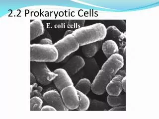
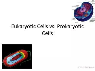
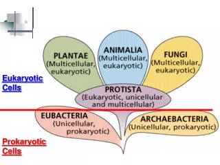
Prokaryotic Cells. Bacteria. Classification of Bacteria. We classify, or name, bacteria based on 3 main factors… Size Shape Arrangement We only study a handful of all bacteria within health science!. Bacterial Size.

E N D
Prokaryotic Cells Bacteria
Classification of Bacteria • We classify, or name, bacteria based on 3 main factors… • Size • Shape • Arrangement • We only study a handful of all bacteria within health science!
Bacterial Size • Surface-to-Volume Ratio: The ratio of total surface space compared to the total volume of a given cell. • The smaller the cell, the larger the surface-to-volume ratio. • A larger surface-to-volume ratio helps with quicker nutrient absorption. • Also allows absorbed nutrients to reach cellular organelles faster!
Bacterial Shapes • The shape of a bacteria is the basic shape of an individual bacteria cell, not the colony. • 5 shapes: • Cocci • Bacilli • Vibrios • Spirochetes • Spirillum
Bacterial Shapes • Cocci: Spherical in shape.
Bacterial Shapes • Bacilli: Rod-like.
Bacterial Shapes • Vibrios: Comma-shaped.
Bacterial Shapes • Spirochetes: Corkscrew-shaped.
Bacterial Shapes • Spirillum: Rigid & wavy-shaped.
Bacterial Arrangements • Bacterial Arrangements: Groups of bacterial cells that have divided without complete separation of the cell walls. • Cocci can divide in many different planes to create.. • Diplo- • Strepto- • Staphylo- • Tetrad- • Sarcina- • Bacilli can only divide on one plane, so produce cells that are connected end-to-end or side-by-side.
Bacterial Arrangements • Diplo-: Pairs of bacterial cells. • Strepto-: Chains of bacterial cells. • Staphylo-: Grapelike clusters. • Tetrad-: Cocci in squares of 4. • Sarcina-: Cocci in squares of 8.
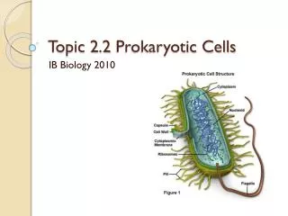
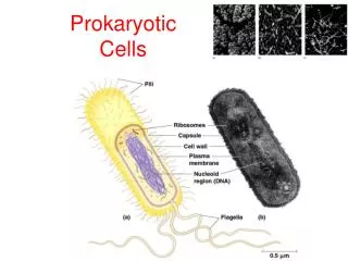
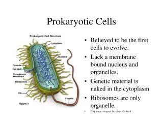
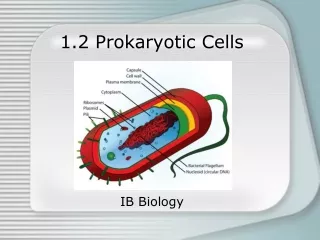
Internal Structures • Cytoplasm: A semifluid substance that makes up the majority of the interior of a bacterium.
Internal Structures • Ribosome: A cellular organelle responsible for translating RNA & synthesizing protein. • Prokaryotic ribosomes are only 70S. • Eukaryotic ribosomes are 80S. • Difference in ribosome sizes aids in chemotherapeutic drugs differentiating between prokaryotic & eukaryotic cells. • Streptomycin & Erythromycin target 70S ribosomes specifically to disrupt protein synthesis & cause cell death only to bacteria.
Internal Structures • Nuclear Region aka Nucleoid: The central region in the bacterial cell where DNA is located. • Bacteria do not have a defined membrane-bound nucleus. • DNA consists of one or more chromosomes, either circular or linear.
Internal Structures • Plasmids: Smaller circular DNA molecules containing genes that supplement the chromosomal material. • Often the site where antibiotic resistant genes are located.
Internal Structures • Inclusions: “Extras” that are not a normal cellular structure. • Granules: Small bodies of densely compacted substances that have not been dissolved in the cytoplasm & without a membrane. • Vesicles: Membrane-bound particles occasionally found that typically play very specialized roles. • Ex. Gas-filled vesicles in cyanobacteria help control their depth in the water.
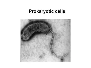
External Structures • Flagella: Long, thin helical appendages that allow motility (movement). • Only 1/10 the thickness of eukaryotic flagella. • About half of known bacteria species are motile. • Basal Region: The point where the flagella attaches to the cell membrane. • Consists of a hook-like structure and a complex basal body. • Basal body consists of a central rod or shaft surrounded by a set of rings. • Gram positive bacteria have one ring in the cell membrane and one in the cell wall. • Gram negative bacteria have a pair of rings in the cell wall.
External Structures • Movement aided by Flagellum: • Chemotaxis: Movement toward or away from a chemical substance in the bacteria’s environment. • Phototaxis: Movement toward or away from light in the bacteria’s environment.
External Structures • Pilli: Tiny hollow projections not involved in movement. • Conjugation Pilli: Pilli designed to attach two bacterial cells together to facilitate the movement of genetic material. Only found in certain groups of bacteria. • Conjugation allows bacteria to pass on antibiotic resistant genes to each other! • Attachment Pilli: Pilli designed to help the bacteria attach to surfaces. • Contributes to pathogenicity of bacterial species since it can aid in attachment to water & air (transfer) and the surface of cells (virulence).
Other Structures • Endospore: A small, compact, tough, structure produced by some bacteria as a means of preserving their genetic material. • NOT a form of reproduction. • Produced when the bacteria’s environment becomes unfavorable or too harsh for survival. • Can remain dormant but viable for thousands of years. • Will germinate (develop) into functional cells once environmental conditions improve. • Most often found in soil & water.
Glycocalyx • Glycocalyx: External structure of polysaccharides or polypeptides outside the cell membrane. • Capsule: Glycocalyx layer that helps protect the bacterium & keep it from being phagocytized. • Considered a virulence factor. • Slime Layer: Glycocalyx layer that is thinner & less tightly bound to the cell wall. • Prevents the cell from drying out. • Helps to trap nutrients near the cell. • Can help cells stick together. • Can help bacteria adhere to objects in the environment. • Plaque on teeth is the slime layer of Streptoccus mutans and a few anaerobes (Fusobacterium & Actinobacteria).
Cell Walls • Cell Wall: The structure that allows us to characterize bacteria into different groups. • Not found in eukaryotic cells, so typically targeted by antibiotics. • Two Important Rolls: • Maintains the characteristic shape of a bacterial cell – would be spherical without it. • Prevents the cell from lysing (bursting) when osmosis triggers fluid to flow into the cell.
Cell Wall • Peptidoglycan: Covalently-linked polymer that surrounds the cell. • Resembles a chain-link fence or net. • Provides support to the cell. • Does not play a major role in regulating entry of materials into the cell. • The most important component of the bacterial cell wall.
Cell Membrane • Cell Membrane: The boundary between the interior of the cell & the environment. • Made up of a lipid bilayer interspersed with proteins. • Some proteins act as carriers, pores, or channels. • Material exchange for the cell occurs through these proteins constantly. • Material exchange for the cell occurs selectively through the lipid bilayer.
Cell Membrane • The primary membrane accessible to the interior of the cell to carry out specialized tasks. • Eukaryotic cells have multiple membrane-bound organelles for this. • Proteins in the cell wall aid in specialized task, including… • DNA replication • Respiration • Cell wall component synthesis • Some proteins are located on the outer surface of the cell membrane. • Includes the proteins that identify the bacteria as a particular organism.
Internal Membrane Systems • Internal membrane Systems: Found in some bacteria, mostly those with photosynthetic capabilities. • Chromatophores: A system of membranes derived from the cell membrane that makes photosynthetic reactions possible.
Differentiating Bacteria • Differentiating bacterial species depends on the cell walls. • Bacteria are named after the staining properties and tests used to “see” them under microscopes. • The different “classes” of bacteria respond in noticeably different ways to these tests.
Gram Positive Bacteria • Gram Positive Bacteria: Bacteria that turn purpleduring a Gram stain. • Very thick peptidoglycan cell wall up to 40 layers thick, with little space between the cell wall & cell membrane. • 60-90% peptidoglycan. • Tiechoic Acid: A unique form of acid in the cell wall that helps set Gram+ bacteria apart. • Periplasm: Fluid located within the network of the cell wall where digestive enzymes are located. • Enzymes destroy substances that are potentially harmful to the bacteria. • Also contains transport proteins to aid in transporting metabolites & nutrients into the cytoplasm.
Gram Negative Bacteria • Gram Negative Bacteria: Bacteria that turn red during a Gram stain. • A bacterial cell with a different external system structure than Gram Positive. • Cell wall is thinner than in Gram+ and more complex. • 10-20% peptidoglycan. • The rest is made up of polysaccharides, proteins, & lipids. • Outer Membrane: Forms the outermost layer on the cell wall, attached to the peptidoglyccan in the cell wall by a layer of small lipoprotein molecules. • Does not control the movement of substances through the cell wall but can control the transport of certain proteins. • Endotoxin: A base of lipopolysaccharides within the outer membrane that is released when the bacterial cell is killed. • This causes fever, vasodilation, & a drop in blood pressure. • Antibiotics given too late in an infection can cause massive release of endotoxins & potentially death. • Periplasmic Space: Space between the cell wall & membrane – takes on the same tasks as the periplasm in G+.
Acid-Fast Bacteria • Acid-Fast Bacteria aka Mycobacteria: Bacteria that does not Gram stain, so is visualized using the Ziehl-Neelsen Acid-Fast Stain. • Cell wall is thick, similar to Gram+ bacteria. • Has less peptidoglycan. • 60% of cell wall is composed of lipids. • Image: A = Non Acid-Fast bacteria, B = Acid-Fast Bacteria
Mycoplasma • Mycoplasma: Bacterium with no cell wall. • Protected from osmostic swelling & lysing by a strong cell membrane. • Cell membrane contains sterols, which provide rigidity – more common in Eukaryotic cells. • More resistant to antibiotic treatment since antibiotics typically attack the cell wall.
Binary Fission • Binary Fission: The method of asexual reproduction used by most prokaryotic cells. • DNA of the mother cell replicates & joins into circular pairs. • The circular pairs attach to the cell membrane/plasma membrane. • The cell elongates, forcing the paired chromosomes separate. • The cell membrane invaginates (pinches inward toward the middle). • When the cell membrane has completed invaginating, the cell splits off into two identical daughter cells. • It is fairly common for one of the daughter cells to not be identical to the mother cell – this causes the high mutation rate of bacteria.
Bacterial Growth Phases • We grow bacteria in nutrient-rich medium (typically broth or agar) in order to watch them grow! • Standard Bacterial Growth Curve: The rate through which bacteria go through the 4 phases of bacterial growth. • Lag Phase • Log Phase • Stationary Phase • Decline Phase • Some bacteria complete this in a few days, some can take years to complete!
Bacterial Growth Phases • Lag Phase: The initial phase where the bacterium is adapting to the environment, particularly if it was previously in a poor environment and is now in an optimal environment. • Can last one hour to several days, depending on previous environment & the need for adaptation. • Bacteria are not currently dividing but are gearing up for it. • Cells increase in size & produce large amounts of ATP energy to prepare.
Bacterial Growth Phases • Log Phase: Bacteria have adapted to the environment and population growth begins. • Growth occurs at a logarithmic rate – this means an exponential and rapid rate of growth. • Generation Time: The genetically determined, rapid rate at which bacteria reproduces. • The generation time is the amount of time it takes for the bacterial population to double.
Bacterial Growth Phases • Stationary Phase: The bacteria have stopped exponential growth and is simply being maintained. • The larger the number of organisms, the faster nutrients are used up & metabolic wastes build up in the environment. • Living space becomes scarce. • The growth curve levels off. • New cells are produced at the same rate as the death of old cells. • The colony does not grow or decline, it remains at a constant rate. • Chemostat: A device used to constantly refresh the medium a bacterial culture is being grown in – prevents stationary phase from occurring.
Bacterial Growth Phases • Decline Phase: The phase at which the bacterial colony begins to die off. • Environmental conditions become increasingly less favorable. • Toxic waste products build up. • Nutrients dwindle. • Cells loose their ability to divide & finally die off. • The number of cells decrease at a rapid logarithmic rate.
Bacterial Environments • Bacteria are found in nearly every environment on earth. • They can be found in places where no other living organism can survive. • This is due to… • Small size • Easily dispersed • Occupy very little space • Need only small quantities of nutrients • Very diverse in nutritional requirements • Live mostly in water. • Can adapt to conditions we would find unpleasant.
Important Environmental Factors • Bacterial Growth Rates can be affected by several environmental factors… • pH • Temperature • Oxygen Content • Moisture • Hydrostatic Pressure • Osmotic Pressure • Nutritional Factors
pH • pH: The measure of the acidity or alkalinity of a substance. • Optimum pH: The pH level at which a bacteria grows best. • Neutrophile: Any bacterium whose optimum pH is neutral, or 5.4-8.0. • Acidophile: Any bacterium whose optimum pH is acidic, or 1.0-5.4. • Alkaliphile: Any bacterium whose optimum pH is alkaline, or 7.0-11.5.
Temperature • Temperature: The measure of warmth or coolness of a substance or the environment itself. • Optimal Temperature: The temperature at which a bacteria grows best. • Mesophile: Any bacterium that grows best at a “warm” temperatures – 25-40°C/77-104°F. • Most common type – includes most human pathogens! • Psychrophile: Any bacterium that grows best at “cold” temperatures – 15-20°C/59-68°F. • Thermophile: Any bacterium that grows best at “hot” temperatures – 50-60°C/122-140°F.
Oxygen • Oxygen: A gaseous element common to many metabolic processes of living things. • Aerobe: A bacterium that requires oxygen for metabolic functions. • Anaerobe: A bacterium that does not require oxygen for metabolic processes.
Oxygen • Obligate Aerobe: A bacterium that MUST have free oxygen present for aerobic respiration – cannot perform anaerobic respiration. • Obligate Anaerobe: A bacterium that cannot tolerate any oxygen in the environment – they use a different molecule for respiration. • Aerotolerant Anaerobe: A bacterium that can survive in the presence of oxygen but do not use it for metabolism. • Microaerophile: A bacterium that grows best in the presence of small amounts of oxygen. • Facultative Anaerobe: A bacterium that carries on aerobic metabolism when oxygen is present but shifts to anaerobic metabolism if oxygen is absent.
Moisture • Moisture: Any liquid required for survival or produced as a metabolic byproduct. • Actively metabolizing bacteria typically require a water-based environment to survive. • Most bacterium can survive a few hours without moisture. • Only spore-forming bacteria can exist in a dormant state in a dry environment.
Hydrostatic Pressure • Hydrostatic Pressure: The pressure exerted by standing water. • The deeper the water, the higher the hydrostatic pressure. • Some bacteria MUST have high hydrostatic pressure. • Ex. Those that live at the bottom of the ocean must have this high pressure for their membranes and enzymes to function properly.
Osmotic Pressure • Osmotic Pressure: The pressure exerted within a solution containing dissolved substances (solutes) within a liquid (solvent). • Osmosis: The diffusion of water through a selectively permeable membrane from the area of higher water concentration to the area of lower water concentration. • The higher solvent (water) concentration area has a low solute concentration. • The higher solute concentration has a low solvent concentration.
Osmotic Pressure • Tonicity: The ability of a solution to affect the fluid volume and the pressure in a cell. • If a solute cannot pass through a plasma membrane, but remains more concentrated on one side of the membrane than on the other, it triggers osmosis. • Hypotonic Solution: Area surrounding a cell has a lower concentration of nonpermeating solutes than the intracellular fluid. • Cells absorb water, swell, and lyse (burst). • Hypertonic Solution: Area surrounding cell has a higher concentration of nonpermeating solutes than the intracellular fluid. • Plasmolysis: The shrinking of a cell in as hypertonic solution due to water loss. • Isotonic Solution: The area surrounding the cell has the same total concentration of nonpermeating solutes as the intracellular fluid. • Cells will neither loose nor gain water molecules & do not change size or shape.
Osmotic Pressure • Halophile: A bacterium specifically designed to require moderate to large quantities of sodium chloride (“salt”) in their environment. • These guys love hypertonic solutions!