Download

1 / 16
170 likes | 641 Views
Modeling Plasmid Selection Joy Killough RET Teacher University of Texas at Austin With Dr. C. Randall Linder Environmental Science Institute Dr. Jay Banner, Director A Research Experience for Teachers Lesson Program Funding from National Science Foundation Modeling Plasmid Selection

E N D
Modeling Plasmid Selection Joy Killough RET Teacher University of Texas at Austin With Dr. C. Randall Linder Environmental Science Institute Dr. Jay Banner, Director A Research Experience for Teachers Lesson Program Funding from National Science Foundation
Modeling Plasmid Selection • Bacteria are very useful organisms in genetic engineering. They are able to bring in plasmids into which DNA of interest may have been added. • Being able to find bacteria containing the plasmid that has been modified requires some techniques that look for the effects of plasmid insertion, which are visible to the naked eye, rather than looking for the plasmid itself (which of course is not). • This activity models the techniques by which bacteria containing modified plasmids can be identified.
Materials Needed For each student group provide • Four ½ sheets of paper cut into ovals to represent bacteria • Four rings of pony beads, each on a full chenille stem (pipe cleaner), formed into a circle to represent a chromosome. Group several beads of a color together to represent genes. • Three smaller rings of beads (half of a stem) to represent plasmids. Make them identical and be sure they have a pair of purple beads (antibiotic resistance gene). Let the first and last bead be green ( lac Z’ gene) and close into a circle leaving an inch or so of the stem at each end without beads. See detailed instructions. • 2 pink beads representing the foreign gene of interest that is being added to the plasmid.
Restriction Enzymes • Restriction enzymes cut DNA at very specific locations. They are predictable, each enzyme always cutting the same way. This characteristic is used in genetic engineering. • Plasmids are cut with the same restriction enzyme used to cut the DNA to be inserted. A restriction enzyme which leaves overhanging sticky ends is needed for this this procedure. This provides the free base pairs needed to combine the plasmid DNA with the source DNA. Restriction Enzyme Cut from EcoRI
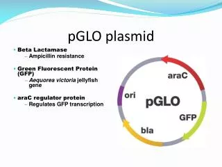
Modeling the plasmid To represent a plasmid, make a small ring of about 10 pony beads strung on a chenille stem (pipe cleaner). Include 2 purple beads to represent the antibiotic resistance gene (ampr) and 2 green beads on each side of the closure to represent the gene Lac Z’. Lac Z’ codes for beta galactosidase, the protein that can cleave the sugar, X-gal, to make a blue colony. Each group of beads of a single color represents a gene. Gene for antibiotic resistance LacZ’ Restriction site Tails of chenille stems
Provide three “plasmid” bead rings and two DNA pieces (pink beads) per student group. Instruct students to add the genes to the plasmids at the restriction site. It is deliberate that they have fewer “gene” beads than plasmids.
Modeling the plasmid cut with restriction enzymes Tell students to open the plasmid by untwisting the chenille stem. This represents the recognition site for the restriction enzyme. Add a pink bead to represent the gene of interest that is now being added to the plasmid since it shares the same sticky ends as the plasmid. Gene for antibiotic resistance Restriction Site LacZ’ Tails of chenille stems
Cut 4 ovals the size of a half a sheet of paper. Each represents a single bacterial cell. Have each student group lay these out on a large surface. Prepare 4 large rings of pony beads strung on pipe cleaners (chenille stems). Sections of beads of a single color represent various genes and the entire ring represents the prokaryotic chromosome. Students should place the chromosome inside the bacterial cell.
Tell students to imagine the area around the bacteria is a Petri dish with nutrient agar. Ask them: If each of these four bacteria were placed on the Petri dish how many would live ? Student should answer all four. Ask them: What would the plate look like? Students should realize each bacteria would form a small white colony. Point out this contains thousands and thousands of bacteria, clones of the original.
Transformation To model transformation have students place their three plasmids into the bacteria, two modified and one original. One bacterium will not receive a plasmid. Ask them: Which bacteria would survive on a plate of nutrient agar? Again they should choose all four.
Selection for Antibiotic Resistance Now have students imagine the area around the bacteria is a Petri dish of nutrient agar with ampicillin and the sugar X-gal added. Ask them: If each of these four bacteria were placed on the this Petri dish how many would live ? This time they should choose three. Only the bacteria with the antibiotic resistance gene (purple) in the plasmid present will survive to form a colony.
Selection for breakdown of X-gal Of the surviving colonies which will be white and which blue? Ask the students why. Answer: The insertion of the pink gene (the gene of interest) into the green LacZ’ gene “breaks” the gene so it can no longer cleave X-gal forming the blue colony. Which colony contain the inserted gene pieces? The white colonies. ampr lacZ’
Modeling Plasmid SelectionStudent Questions • 1. How are prokaryotic chromosomes and eukaryotic chromosomes different? • 2. What is a plasmid? • 3. How is a plasmid different from a chromosome? • 4. Why are indirect means used to identify the presence of an inserted piece of DNA in a plasmid? • 5. How do restriction enzymes cut? • 6. What are sticky ends and why are they so important? • 7. What procedure makes it likely that a plasmid and a piece of foreign DNA will combine? • 8. Research the restriction enzyme and find the difference between a blunt cut and a cut which leaves sticky ends. • 9. Research the restriction enzyme and find out how they are named. • 10. Research the discovery of restriction enzymes.
Modeling Plasmid Selection Answers • 1. How are prokaryotic chromosomes and eukaryotic chromosomes different? One difference is that prokaryotic cells typically have a single circular chromosome whereas eukaryotic cells have linear, paired chromosomes. • 2. What is a plasmid? A plasmid is an extra, circular, piece of DNA outside of the bacterial chromosome which replicates independently. • 3. How is a plasmid different from a chromosome? Plasmids are smaller than chromosomes and contain genes that are normally not critical to the functioning of the cell. • 4. Why are indirect means used to identify the presence of an inserted piece of DNA in a plasmid? Plasmids and inserted genes are too small to see. • 5. Where do restriction enzymes cut? Restriction enzymes recognize a specific sequence of DNA called a recognition sequence. Each restriction enzyme cuts at a particular restriction site where a recognition sequence is found.
6. What are sticky ends and why are they so important? Sticky ends are overhanging single strands of DNA left behind from restriction enzyme cuts. The sticky ends provide a way for a piece of foreign DNA to be inserted following the base pairing rules. • 7. What procedure makes it likely that a plasmid and a piece of foreign DNA will combine? Cutting the plasmid and the piece of DNA to be inserted with the same restriction enzyme gives cuts with the same sticky ends allowing for easy combining. • 8. Research the restriction enzyme and find the difference between a blunt cut and a cut which leaves sticky ends. Answers will vary, but should include that blunt cuts cut straight through both strands of DNA at the same location and that sticky ends leave a single stranded overhang. • 9. Research the restriction enzyme and find out how they are named. Answers will vary, but should include that they are named using the genus, species and strain of the bacterium they are recovered from as well as a number reflecting the order of identification from that bacterium. • 10. Research the discovery of restriction enzymes. Answers will vary.
References • Campbell, Neil, and Jane Reece. Biology. 6th ed. San Francisco, California: Benjamin Cummings, 2002. • Raven, Peter, and George Johnson. Biology. 6th ed. Boston, Massachusetts: McGraw-Hill, 2002.