Download
1 / 66
710 likes | 1.66k Views
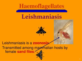
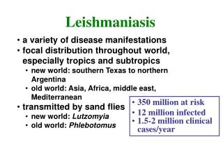
Haemoflagellates Leishmaniasis & Trypanosomiasis. Different stages of Haemoflagellates. Promastigotes of Leishmania . Amastigote of Leishmania. The life cycle of Leishmania. Leishmania Parasites and Diseases. World distribution of Visceral Leishmaniasis. Sand fly.

E N D
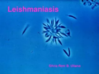
Promastigotes of Leishmania Amastigote of Leishmania
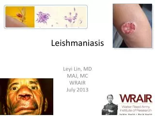
Clinical types of cutaneous leishmaniasis • Leishmania major:Zoonotic cutaneous leishmaniasis: wet lesions with severe reaction • Leishmania tropica:Anthroponotic cutaneous leishmaniasis: Dry lesions with minimal ulceration Oriental sore (most common) classical self-limited ulcer
Uncommon types • Diffuse cutaneous leishmaniasis (DCL): Caused by L. aethiopica, diffuse nodular non-ulcerating lesions. Low immunity to Leishmania antigens, numerous parasites. • Leishmaniasis recidiva (lupoid leishmaniasis): Severe immunological reaction to leishmania antigen leading to persistent dry skin lesions, few parasites.
Diffuse cutaneous leishmaniasis Leishmaniasis recidiva
cutaneous leishmaniasis Diagnosis: • Smear: Giemsa stain – microscopy for LD bodies (amastigotes) • Biopsy: microscopy for LD bodies or culture in NNN medium for promastigotes
Treatment • No treatment – self-healing lesions • Medical: • Pentavalent antimony (Pentostam), Amphotericin B • +/- Antibiotics for secondary bacterial infection. • Surgical: • Cryosurgery • Excision • Curettage
Pentostam ( sodium stibogluconate) for treatment of all types of leishmaniasis
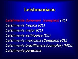
Visceral leishmaniasis • There are geographical variations. • The diseases is called kala-azar • Leishmania infantum mainly affect children • Leishmania donovani mainly affects adults
Presentation • Fever • Splenomegaly, hepatomegaly, hepatosplenomegaly • Weight loss • Anaemia • Epistaxis • Cough • Diarrhoea
Untreated disease can be fatal After recovery it might produce a condition called post kala-azar dermal leishmaniasis (PKDL)
Visceral leishmaniasis Diagnosis • Parasitological diagnosis: METHOD Bone marrow aspirate 1. microscopy Splenic aspirate 2. culture in NNNmedium Lymph node Tissue biopsy
Bone marrow aspiration Bone marrow amastigotes
(2) Immunological Diagnosis: • Specific serologic tests: Direct Agglutination Test (DAT), ELISA, IFAT • Skin test (leishmanin test) for survey of populations and follow-up after treatment. • Non specific detection of hypergammaglobulinaem by formaldehyde (formol-gel) test or by electrophoresis.
DAT test ELISA test
Treatment: • Pentavalent antimony- sodium stibogluconate (Pentostam) • Amphotericin B Treatment of complications: • Anaemia • Bleeding • Infections etc.
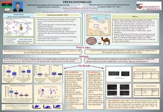
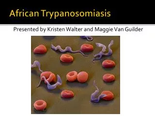
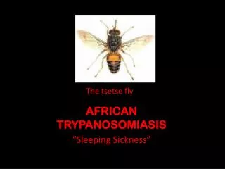
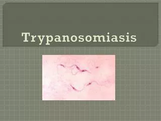
African Trypanosomiasis Life cycle of Trypanosoma brucei gambiense & T. b. rhodesiense
African sleeping sickness Trypanosoma brucei rhodesiense: East Africa, wild and domestic animal reservoirs Trypanosoma brucei gambiense: West and Central Africa, mainly human infection
Pathology and clinical picture • Skin stage: chancre. • Haematolymphatic stage: generalized lymphadenopathy, anaemia, generalized organ involvement. • Central nervous system stage (CNS): Meningoencephalitis. (Development of the disease more rapid in Trypanosoma brucei rhodesiense)
AMERICAN TRYPANOSOMIASIS LIFE CYCLE OF Trypanosoma cruzi